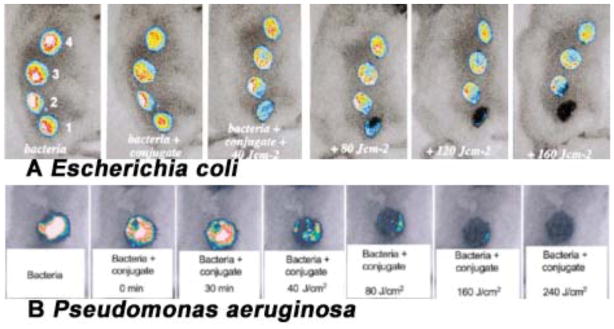
Fig. (12). PDT for infected excisional wounds.
(A) Successive overlaid luminescence images of a mouse with four excisional wounds infected with equal numbers of E. coli (5 ×10⁶). Wounds 1 (nearest tail) and 4 (nearest head) received topical application of pL-ce6 conjugate 22. Wounds 1 and 2 (two nearest tail) were then illuminated with successive fluences (40–160 J/cm2) of 665 nm light. (B) Successive overlaid luminescence false-color images of mice bearing excisional wounds infected with 5×10⁶ luminescent P. aeruginosa treated with pL-ce6 conjugate 22 and increasing doses of 660 nm light.

