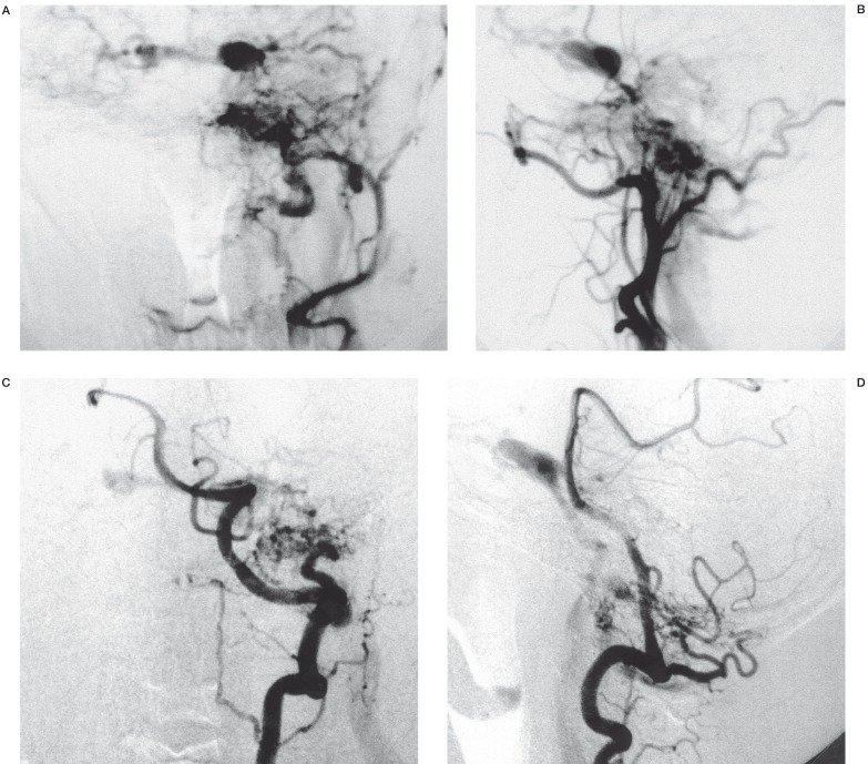
Figure 2.
Anteroposterior (A) and lateral projection (B) of Left external carotid arteriogram. Anteroposterior (C) and lateral projection (D) of left VA arteriogram. DAVF was supplied by meningeal branches of the left ascending pharyngeal artery and vertebral artery and drained the left internal jugular veins and IPS, then to the bilateral CS and cortical veins.

