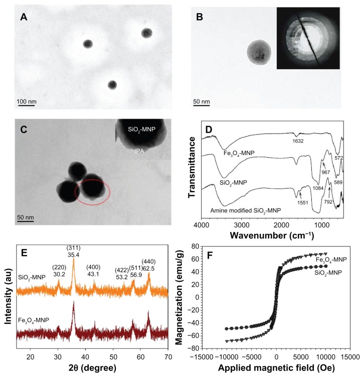
Figure 3.
Transmission electron microscope images of (A and B) silica-coated magnetic nanoparticle (SiO2-MNP) and (C) tissue plasminogen activator (tPA) bound to SiO2- MNP (SiO2-MNP-tPA) after staining with phosphotungstic acid. Characterization of superparamagnetic iron oxide magnetic nanoparticle (Fe3O4-MNP) and SiO2-MNP with (D) Fourier transform infrared spectroscopy analysis, (E) X-ray diffraction patterns, and (F) superconducting quantum interference device magnetization curves.
Notes: (A) Magnification = 100,000, bar = 100 nm; (B and C) magnification = 300,000, bar = 50 nm. Insert in (B) is electron diffraction pattern; insert in (C) is a blowup of the circled area.

