ABSTRACT
In eukaryotes, DNA synthesis is preceded by licensing of replication origins. We examined the subcellular localization of two licensing proteins, ORC2 and MCM7, in the mouse zygotes and two-cell embryos. In somatic cells ORC2 remains bound to DNA replication origins throughout the cell cycle, while MCM7 is one of the last proteins to bind to the licensing complex. We found that MCM7 but not ORC2 was bound to DNA in metaphase II oocytes and remained associated with the DNA until S-phase. Shortly after fertilization, ORC2 was detectable at the metaphase II spindle poles and then between the separating chromosomes. Neither protein was present in the sperm cell at fertilization. As the sperm head decondensed, MCM7 was bound to DNA, but no ORC2 was seen. By 4 h after fertilization, both pronuclei contained DNA bound ORC2 and MCM7. As expected, during S-phase of the first zygotic cell cycle, MCM7 was released from the DNA, but ORC2 remained bound. During zygotic mitosis, ORC2 again localized first to the spindle poles, then to the area between the separating chromosomes. ORC2 then formed a ring around the developing two-cell nuclei before entering the nucleus. Only soluble MCM7 was present in the G2 pronuclei, but by zygotic metaphase it was bound to DNA, again apparently before ORC2. In G1 of the two-cell stage, both nuclei had salt-resistant ORC2 and MCM7. These data suggest that licensing follows a unique pattern in the early zygote that differs from what has been described for other mammalian cells that have been studied.
Keywords: chromatin, DNA replication, early development, fertilization
The DNA replication licensing protein MCM7 binds to DNA before origin recognition complex, subunit 2 (ORC2), and ORC2 appears to play significant roles in chromatin organization and nuclear structure.
INTRODUCTION
DNA replication in eukaryotic cells requires multiple origins of replication. In mammals, there are on the order of 103 to 105 origins per cell that are required to efficiently replicate the genome [1, 2]. These origins fire at different times during S-phase, which poses a problem for the cell—how to ensure that each origin initiates DNA replication only once so that the entire genome is replicated without duplications. Blow and Laskey [3] proposed and then provided experimental evidence [4, 5] for a mechanism called licensing through which each origin is made competent for replication in G1. Licensing occurs when a series of proteins, beginning with the origin recognition complex proteins (ORC1-6) and ending with the helicases MCM2-7, bind to origins during the M-to-G1 transition [6–9]. In mammalian somatic cells, ORC2-5 remain associated with origins during the entire cell cycle [6, 7, 10, 11]. ORC1 loads onto this complex, and the binding of MCM2-7 complex completes licensing of the origin. New licensing is restricted from occurring in S-phase, and only licensed origins are replicated.
Mammalian sperm and oocyte chromatin have several unique aspects that prevent the formulation of an immediately obvious hypothesis as to when licensing proteins, particularly ORC2-5, are recruited to the origins. Both gametes are the products of meiosis, not mitosis, and may therefore not retain ORC2-5 as do cycling somatic cells [6, 7, 10, 11]. Moreover, the very tightly packaged sperm chromatin [12–14] would not be expected to carry licensing proteins. In support of this, we have demonstrated that paternal pronuclei resulting from the injection of sperm halos that had been extracted with 2 M NaCl are competent for DNA replication [15]. Since the known licensing proteins cannot remain bound to DNA under this condition, the experiment suggests that all licensing proteins can be supplied by the oocyte. This suggests that for the zygote, licensing may be de novo, beginning with the association of ORC2-5.
The zygote is also unique in that two different pronuclei replicate DNA in the same cytoplasm [16], posing questions about the different conditions for licensing in each pronucleus. After fertilization, sperm chromatin must decondense before the protamine-bound DNA becomes accessible to licensing proteins [17]. However, some studies have suggested that licensing can occur as early as during mitosis [18], so the maternal DNA is theoretically accessible to licensing proteins even before fertilization. Sperm that are injected into oocytes up to 3 h after parthenogenetic activation can still replicate DNA [19, 20]. Thus, licensing may be asynchronous in the two pronuclei.
Because of these unique aspects of zygotic DNA replication, we have initiated a study of licensing in the mouse one-cell embryo. We began by investigating one of the first licensing proteins known to adhere to the origin, ORC2, and one of the last, MCM7. Our data demonstrate that the maternal and paternal chromatin are licensed asynchronously and that some patterns of the appearance of these licensing proteins on the DNA are different from what has been reported for somatic cells with MCM7 appearing on the DNA before ORC2.
MATERIALS AND METHODS
Animals
B6D2F1 (C57BL/6N X DBA/2) mice were obtained from National Cancer Institute (Raleigh, NC). Mice were kept in accordance with the guidelines of the Laboratory Animal Services at the University of Hawaii and those prepared by the Committee on Care and Use of Laboratory Animals of the Institute of Laboratory Resources National Research Council (DHEF publication no. [NIH] 80-23, revised 1985). The protocol for animal handling and the treatment procedures were reviewed and approved by the Institutional Animal Care and Use Committee at the University of Hawaii.
Collection of Gametes
The luminal content of the caudal epididymides and vas deferens of ∼8-wk-old mice was extracted separately and suspended in HCZB [21]. Mature females, 8–12 wk old, were induced to superovulate with i.p. injections of 5 IU eCG and 5 IU hCG given 48 h apart. Oviducts were removed 14–15 h after the injection of hCG and placed in HCZB. The cumulus-oocyte complexes were released from the oviducts into 0.1% of bovine testicular hyaluronidase (300 USP units/mg) in HCZB medium to disperse cumulus cells. The cumulus-free oocytes were washed with HCZB medium and used immediately for injection.
Intracytoplasmic Sperm Injection
Spermatozoa were prepared for Intracytoplasmic sperm injection (ICSI) by mixing with 12% w/v polyvinyl pyrolidone (PVP; 360 kDa); single sperm heads were injected into each oocyte. ICSI was carried out as described by Szczygiel and Yanagimachi [21]. A small drop of treated sperm suspension was mixed thoroughly with an equal volume of HCZB containing 12% (w/v) (Mr 360 kDa) immediately before ICSI. ICSI was performed using Eppendorf Micromanipulators (Micromanipulator TransferMan, Eppendorf, Germany) with a piezoelectric actuator (PMM Controller, model PMAS-CT150; Prime Tech, Tsukuba, Japan). A single spermatozoon was drawn, tail first, into the injection pipette and moved back and forth until the head-midpiece junction (the neck) was at the opening of the injection pipette. The head was separated from the midpiece by applying one or more piezo pulses. After discarding the midpiece and tail, the head was redrawn into the pipette and injected immediately into an oocyte. After ICSI, oocytes were cultured in CZB [22] for 5 h at 37°C in 5% CO2 in air.
Antibodies
Polyclonal goat anti-ORC2 (C-18, catalog no. sc-13238; Santa Cruz Biotechnology, Santa Cruz, CA) and monoclonal mouse-MCM7 (G-7, catalog no. 46687; Santa Cruz Biotechnology) primary antibodies were used. We used a mouse monoclonal antibody that recognizes both TUBG1 and TUBG2 (catalog no. sc-17787; Santa Cruz Biotechnology). We have referred to the protein as TUBG in this article to denote that we are cannot be certain which protein the antibody recognized. Secondary antibodies included Alexa Fluor 488 donkey anti-rabbit, Alexa Fluor 488 rabbit anti-goat, and Alexa Fluor 546 rabbit anti-mouse (Invitrogen, Grand Island, NY). The Click-iT EdU Alexa Fluor 488 HCS Assay kit was used according to the manufacturer's protocol (catalog no. C10350; Invitrogen) to verify entry into S-phase.
Immunocytochemistry
Cells were cultured in CZB and fixed in 4% paraformaldehyde for 30 min at room temperature. Paraformaldehyde stock solutions were prepared by diluting a 16% stock solution (Alfa Aesar stock no. 43368) to 4% then stored at 4°C overnight prior to fixing. After fixing, cells were rinsed once and washed twice in 0.1% Tween/PBS for 10 min (PBSw). Cells were permeabilized in 0.5% Triton X-100 for 15 min, after which they were rinsed once and washed twice in PBSw with 0.5% BSA. Cells were blocked with BSA stock for 1 h at room temperature, then incubated in primary antibody at 1:300 dilution overnight at 4°C. Cells were rinsed once and washed twice in washing media, then incubated in secondary antibody at 1:1000 dilution at room temperature for 1 h. Cells were again rinsed and washed in washing media then mounted with ProLong Gold antifade reagent with DAPI (catalog no. P-36931; Invitrogen). Cells were analyzed with an FV1000-IX81 confocal microscope from Olympus using Fluoview v. 2.1 software.
Cell Extraction
To observe DNA-bound ORC2 and MCM7 within pronuclei of the one-cell embryo and nuclei in the two-cell embryo, nuclear membranes were permeabilized and washed according to a modified method used by Swiech et al. [23]. Embryos were washed in ice-cold Ca+2/Mg+2-free PBS containing 0.5 mM PMSF, 10 μg/ml of leupeptin and aprotinin (modified ice-cold cytoskeleton buffer, mCSK) for 3 min on ice followed by washing in mCSK with 0.5% Triton X-100, and 100 mM NaCl for 2 min on ice, then a final wash was performed in mCSK for 3 min on ice. Cells were then fixed according to the immunocytochemistry protocol stated.
Analysis of ORC2 and MCM7 Localization
For each stage of development of the zygote and two-cell embryo that was analyzed, at least three separate experiments were performed on different days with each experiment having at least 30 embryos per group. The image chosen was representative of observed localization. Figures 2, 3, and 5 and Supplemental Figures S2 and S3 show cells from a single series, so their intensities are relative.
RESULTS
ORC2 First Appears Adjacent to the Maternal DNA Just after Fertilization
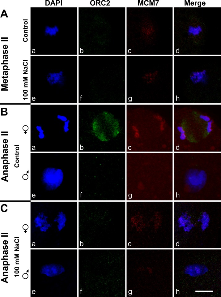
The mouse oocytes are arrested in metaphase of the second meiotic division (MII). MII oocytes do not contain detectable ORC2 (Fig. 1A, b and f). Within an hour after fertilization, the maternal chromosomes go through anaphase to release the second polar body (anaphase II). We found that ORC2 appeared in the area between the separating chromosomes of the metaphase plate in two distinct clusters of foci that paralleled the chromosomes (Fig. 1Bb). Both MCM7 and ORC2 remain associated with the nucleus when it is extracted with 100 mM NaCl only if they are loaded onto the DNA [24]. At this stage, ORC2 did not appear to be directly associated with DNA at all and was largely salt extractable (Fig. 1Bf). A similar pattern for ORC2 localization was previously noted in HeLa cells, except that ORC2 was also present on the mitotic centromeres [24].
FIG. 1.
Localization of ORC2 and MCM7 in the zygote 1–2 h after fertilization. MII oocytes or zygotes shortly after fertilization were fixed and double immunostained with antibodies to ORC2 (green, b and f) and MCM7 (red, c and g), and counterstained with DAPI (a and e), then visualized by confocal microscopy. For clarity, the images in this figure have been enlarged to show only the chromatin and pronuclei. A) Metaphase II oocytes were not treated (Aa–Ad) or extracted with 100 mM NaCl to remove soluble ORC2 and MCM7 (Ae–Ah). B) A zygote shortly after fertilization that was not extracted with salt. The maternal chromosomes are separating in anaphase II. Ba–Bd) Confocal planes at the separating chromosomes of the maternal anaphase II. Be–Bh) A different set of confocal planes showing the decondensing sperm nucleus of the same zygote shown in Ba–Bd. Note that the paternal DNA has no detectable MCM7 (Bg). C) Anaphase II zygote as in B but extracted with 100 mM NaCl to remove soluble ORC2 and MCM7. Ca–Cd) Maternal DNA. Ce–Ch) Recondensing sperm chromatin. This zygote had progressed slightly farther than the one in B, and now the paternal chromatin had MCM7 (Cg) but not ORC2 (Cf). Bar = 10 μm.
MCM7 Is Detectable on the Chromatin Before ORC2
We found that MII oocytes did contain detectable MCM7 (Fig. 1Ac). We tested whether this MCM7 was bound to DNA by extracting with 100 mM NaCl. In MII oocytes, MCM7 staining was resistant to salt washing (Fig. 1Ag) and remained associated with the maternal chromatin as the chromosomes began to segregate during anaphase II of maternal meiosis (Fig. 1Cc). As noted above, at this time, ORC2 was localized to the point between the separating chromosomes, suggesting that MCM7 associated with DNA before ORC2.
A similar pattern for MCM7 was found for the paternal DNA; however, there was no corresponding ORC2 accumulation adjacent to the chromatin when MCM7 first appeared on the sperm DNA. At about 1.5 h after fertilization, while the maternal nucleus goes through anaphase II, the sperm nucleus has completed its first decondensation/recondensation phase, and the protamines are replaced by histones [25, 26]. Early during this decondensation/recondensation, neither ORC2 nor MCM7 was present on the sperm DNA (Fig. 1B, f and g). Later, MCM7 appeared as foci on the sperm DNA, but ORC2 was not detectable (Fig. 1B, f and g). Salt extraction confirmed that the MCM7 was associated with the paternal DNA (Fig. 1Cg), and ORC2 was still not detectable (Fig. 1Cf and Supplemental Fig. S1Bf; all Supplemental Data are available online at www.biolreprod.org). These data suggest that for both maternal and paternal DNA, MCM7 associates with DNA before ORC2.
Both Pronuclei Contain DNA-Bound ORC2 and MCM7 at 4 h after Fertilization
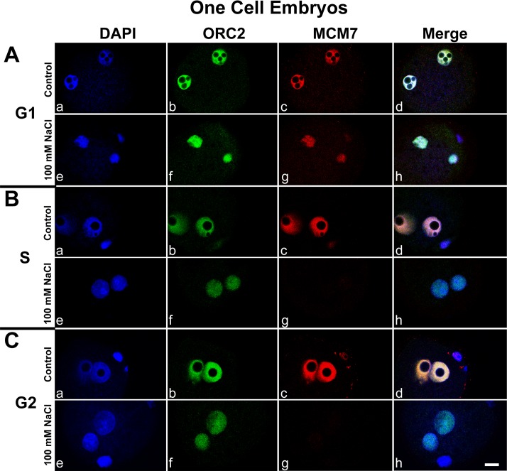
At about 2 h after fertilization, when telophase II was not yet completed, the female pronucleus was positive for both ORC2 and MCM7 (Supplemental Fig. S1B, b and c), while the male pronucleus was positive only for MCM7 (Supplemental Fig. 1B, f and g). By 4 h after fertilization mitosis is complete, the sperm nucleus is loaded with histones, and both pronuclei had formed and are in G1 [25, 26]. At this time point, both ORC2 and MCM7 had accumulated within both pronuclei (Fig. 2A, b and c) and remained associated after salt extraction (Fig. 2A, f and g), suggesting that mouse zygotic pronuclei are licensed for DNA replication by 4 h after fertilization if not before (see Discussion). This is consistent with our previous report showing that pronuclei that were extracted from mouse embryos 4 h after fertilization were capable of DNA synthesis when they were transferred to parthenogenetically activated S-phase oocytes [20]. Since licensing cannot occur in S-phase, we suggested that the transferred pronuclei were already licensed by 4 h. DNA synthesis begins between 5 and 6 h after fertilization whether by in vitro fertilization or ICSI [26], and our data suggest that licensing is complete approximately 2 h before DNA synthesis.
FIG. 2.
Depletion of MCM7 but not ORC2 during zygotic S-phase. A) During late G1, about 4 h after fertilization, both ORC2 and MCM7 remain tightly associated with DNA (Af and Ag). B) During S-phase, soluble ORC2 and MCM7 are present (Bb and Bc), but only ORC2 remains bound after salt extraction (Bf). C) G2 zygotes have the same pattern as S-phase zygotes. Zygotes were fixed and double immunostained with antibodies to ORC2 (green, b and f) and MCM7 (red, c and g) and counterstained with DAPI (a and e), then visualized by confocal microscopy. Zygotes were stained without treatment (a–d) or after extraction with 100 mM NaCl to remove soluble ORC2 and MCM7 (e–h). Bar = 10 μm.
In the First Cell Cycle, DNA-Bound MCM7 Decreases During S-Phase, but DNA-Bound ORC2 Does Not
The first S-phase occurs before the fusion of the pronuclei. We consistently found that MCM7 and ORC2 remained in both pronuclei during S-phase (Fig. 2B, b and c). We confirmed the DNA replication status of these zygotes with EdU staining (Supplemental Fig. S2). However, when these zygotes were extracted with 100 mM NaCl before immunostaining, only ORC2 remained bound (Fig. 2Bf and Supplemental Fig. S2G), while MCM7 was washed out of the pronuclei (Fig. 2Bg and Supplemental Fig. S2O). These data demonstrate that MCM7 remains in the nucleus during S-phase but, as expected, is released from the DNA as S-phase progresses, while ORC2 remained associated with the DNA. ORC2 remained bound to the DNA in G2 (Fig. 2C, b and f), while MCM7 was present only in its soluble form (Fig. 2C, c and g).
ORC2 Is Displaced from the Chromatin During Zygotic Mitosis
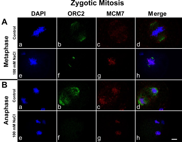
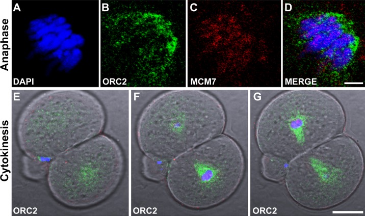
With the onset of the first zygotic mitosis, ORC2 was not detectable in the chromosomes but was present at the spindle poles (Fig. 3Ab). Most of this ORC2 was extractable by salt (Fig. 3Af), though a small residue remained tightly bound, perhaps to the centrosomes (see below). MCM7, however, appeared to be bound to the DNA, as it was still detectable after salt washing (Fig. 3Ag). ORC2 was clearly present in the area between the separating chromosomes and adjacent to the DNA, somewhat similar to its localization in anaphase II of the fertilized oocyte (Figs. 1Bb and 3Bb). Higher-resolution images show that by early anaphase, ORC2 appeared to localize around the separating chromosomes, fitting the spaces in between almost like a glove (Fig. 4B), while MCM7 was localized to the chromatin (Fig. 4C). By cytokinesis, ORC2 surrounded the developing nuclei like a sphere (Fig. 4, E–G). Most of this ORC2 was extractable by salt washing (Fig. 3Bf), suggesting that this localization of ORC2 was not tightly associated with another cellular structure or bound to DNA. Throughout mitosis, MCM7 remained associated with DNA (Figs. 3Ag and 3Bg; see also Supplemental Fig. S3). These data show that two aspects of ORC2 and MCM7 at zygotic mitosis were similar to the behavior of the proteins at the second metaphase of maternal meiosis just after fertilization. First, ORC2 localized at the spindle poles, then adjacent to the separating chromosomes. Second, MCM7 bound the DNA before ORC2.
FIG. 3.
ORC2 and MCM7 localization during zygotic mitosis. A) ORC2 localizes to the spindle poles at zygotic metaphase (Ab), but most of this staining is washed away with salt extraction (Af). MCM7 remains associated with DNA after salt extraction (Ag). B) During anaphase, ORC2 surrounds the developing nuclei (Bb and Bd) while MCM7 is associated with DNA (Bg). Zygotes were fixed and double immunostained with antibodies to ORC2 (green, b and f) and MCM7 (red, c and g) and counterstained with DAPI (a and e), then visualized by confocal microscopy. Zygotes were stained without treatment (a–d) or after extraction with 100 mM NaCl to remove soluble ORC2 and MCM7 (e–h). Bar = 10 μm.
FIG. 4.
High-resolution localization of ORC2 during zygotic mitosis shows that it accumulates around the newly formed nucleus. A-D) High-resolution confocal image of one chromatin set that has already separated from the other in late anaphase. Note that ORC2 appears to surround the chromosomes (B) while MCM7 colocalizes with the DNA (C). Bar in D = 4 μm for A–D. E–G) Three successive confocal planes of a dividing one-cell embryo at cytokinesis showing DAPI (blue) and ORC2 (green). ORC2 appears to surround the chromatin in a sphere in the lower cell. Bar in G = 20 μm for E–G.
ORC2 Localizes Near TUBG Organizing Centers
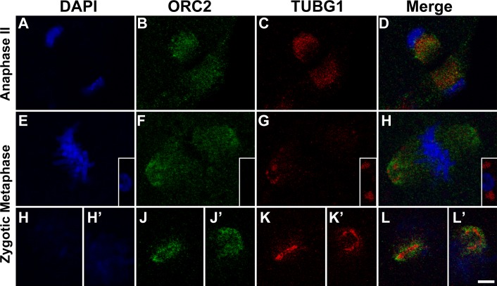
Because we found that ORC2 localized near the spindle poles in zygotic mitosis, we tested whether ORC2 was also localized with TUBG (previously γ-tubulin), a major component of centrosomes. A previous study demonstrated that TUBG is localized between the separating maternal chromosomes of anaphase II, similar to what we have shown for ORC2 (Fig. 1Bb) [27]. These authors also showed that TUBG is diffuse around the mouse zygotic spindle poles and is not limited to tight focus at the centrioles as in somatic cell metaphase plates [24]. We found that TUBG colocalized with the ORC2 that was present between the separating maternal chromosomes in anaphase II shortly after fertilization. Figure 5, B and C, demonstrates that while there is some overlap in the two proteins, ORC2 seems to emanate from TUBG. In the zygotic metaphase, TUBG was localized in a diffuse area at the spindle poles often as a large, ring-like structure (Fig. 5G). TUBG was localized to smaller, tight foci in the polar body mitoses (Fig. 5G, inset). We found that ORC2 was present at the spindle poles (Fig. 5F) and that it seemed to extend from the TUBG1 ring. One other example of the TUBG1/ORC2 colocalization from another zygote at the same stage is shown in Figure 5, H–L′. In this case, it is clear that ORC2 is positioned around a nucleus of TUBG1.
FIG. 5.
Colocalization of OCR2 and TUBG1 in anaphase II and zygotic mitosis. Zygotes were fixed and double immunostained for ORC2 (B, F, J, and J′) and for TUBG1 (C, G, K, and K′) and counterstained with DAPI (A, E, H, and H′). A–D) Anaphase II of the maternal chromosomes shortly after fertilization. ORC2 is localized to two foci between the separating chromosomes, as in Figure 1Bb (B). TUBG1 is closely associated with ORC2 but is closer to the DNA. E–H) ORC2 (F) and TUBG1 (G) are at the spindle poles. TUBG1 appears as a loose ring-like structure (G). The polar body demonstrates a more tightly focused TUBG1 signal, more similar to somatic cells (G, inset). H–L′) The two spindle poles from another zygote. In one spindle pole, TUBG1 appears as a linear signal (K) with ORC2 surrounding the TUBG1 (J), while in the second spindle pole, TUBG1 appears as a ring (K′) with ORC2 surrounding the TUBG1 ring (J′). Bar = 5 μm.
Licensing in the Two-Cell Embryo
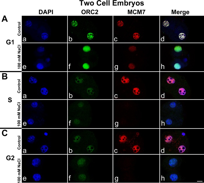
In the two-cell embryo, MCM7 behaved as it did in the zygote, but ORC2 showed a decrease in nuclear staining intensity during S-phase. We found that ORC2 and MCM7 accumulated in the nuclei during G1 of the two-cell embryo, as expected (Fig. 6A, b and c) and that both proteins were resistant to salt extraction (Fig. 6B, f and g). During S-phase, ORC2 remained bound to the DNA but in much diminished amounts (Fig. 6Bf), while MCM7 was released from the chromatin (Fig. 6Bg). MCM7 appeared to remain in the nucleus in its soluble form (Fig. 6Bc), but total ORC2 was diminished (Fig. 6Bb). In G2, MCM7 was present in the nuclei (Fig. 6Cc) but only in its soluble form (Fig. 6Cg). ORC2 appeared to remain bound to DNA though in the same low amounts as was seen in S-phase (Fig. 6C, b and f). These findings were verified by EdU staining.
FIG. 6.
ORC2 and MCM7 during the second embryonic cell cycle. Staining for ORC2 and MCM7 for the two-cell embryo. Panels are arranged exactly as for Figure 2 but at the two-cell stage. Note that ORC2 appears to decrease somewhat during S-phase, but what is left remains resistant to salt extraction (Bb and Bf). All images in this figure were taken on the same day from the same experimental series with the same microscope settings. Images are representative from several experiments. Bar = 10 μm.
DISCUSSION
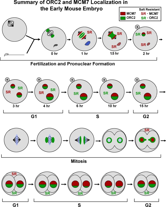
The mouse embryo is a unique model for DNA replication for the reasons already noted in the Introduction. Our data provide several previously undefined aspects of localization of licensing proteins in the first cell cycle. Mouse oocytes contain ORC2-5 [28] and MCM7 [23, 29], so the proteins do not need to be synthesized and are available for deposit onto both parental chromosomes at fertilization. The key differences that we have found between the embryo and what has been reported for somatic cells are that MCM7 binds to the maternal and paternal chromatin of the zygote before ORC2 and that ORC2 forms a ring around the newly forming nuclei of the two-cell embryo. The data are summarized in Figure 7.
FIG. 7.
Summary of ORC2 and MCM7 localization during the first two cell cycles of the mouse embryo. ORC2 is depicted in green, MCM7 is depicted in red, and DNA is depicted in blue. The relative levels of ORC2 and MCM7 within zygotic pronuclei and two-cell embryo nuclei are depicted by the amount of green and red. When ORC2 was salt resistant, a green SR is shown. When MCM7 was salt resistant, a red SR is shown.
ORC2 Localization During Metaphase
Prasanth et al. [24] reported that in HeLa cells, ORC2 localized at the spindle poles during mitosis. This report also included one image in which ORC2 accumulated between the separating chromosomes, as we have shown for the mouse zygote (Fig. 1Cb), but its function here remains unclear. There were two important differences in ORC2 localization between the cycling HeLa cells [24] and mouse zygotes in this study. First, in HeLa cells, ORC2 also localized to the centromeres throughout the cell cycle, while we did not detect ORC2 on mitotic chromosomes. Second, there was a significant level of ORC2 in the cytoplasm of HeLa cells, while we did not detect a significant level of ORC2 in the cytoplasm until the two-cell stage. We suggest that this accumulation of ORC2 that we noted between the separating anaphase II chromosomse serves as a store for its subsequent accumulation in the pronucleus.
ORC2 may play important roles in chromosome segregation in addition to its role in DNA licensing. In this respect, it is interesting to note that ORC2 accumulation was not noted in the paternal chromatin until pronuclear formation, about 4 h after fertilization, emphasizing its potential role in chromatin segregation as opposed to chromatin decondensation. The association of ORC2 with TUBG1 supported such a role. A strong concentration of ORC2 was seen near condensed TUBG1-rich structures, suggesting that the ORC2 was organized by centrioles. In the area between the separating chromosomes, TUBG1 appeared to be more loosely associated with the accumulated ORC2 (Fig. 6D), with little or no structure in this area.
MCM7 Binds to DNA Before ORC2
Our results demonstrate that unlike somatic cells in which the ORC2 binds before MCM7 [6, 7, 10, 11], in the zygote and in the two-cell embryo, MCM7 binds to DNA before ORC2. This is the clearest in the forming paternal pronucleus, which contains MCM7 but no detectable ORC2 (Fig. 1C), but is also convincing for both the maternal chromatin of the zygote (Fig. 1A) and the forming nuclei of the two-cell embryo (Fig. 3A). We cannot say whether MCM7 was loaded on DNA replication origins as a licensing complex, but it does appear to be bound to DNA because it withstands 100 mM NaCl extraction. In most organisms, the licensing is limited to G1 by inhibiting the loading of the MCM helicases to the origins in all other phases of the cell cycle, and the ORC complex appears to direct the MCM2-7 complex to the origins in S-phase [30, 31].
Implications for the Order of ORC2 and MCM7 Binding in Zygotes
Most studies suggest that ORC2 binding to DNA is required for MCM7 loading. But recent publications have suggested that this model is still evolving. Prasanth et al. [24] depleted Orc2 mRNA from HeLa cells and were surprised to find that MCM7 still loaded onto DNA [24]. They were unable to determine if the Orc2 depleted cells could replicate because of technical considerations of the experiment. The authors of another recent study with Caenorhabditis elegans were able to detect ORC2 in the anaphase chromosomes of the fertilized embryo, but their data suggested that the loading of the ORC complex was dynamic, while MCM7 loading was more permanent [32]. They proposed a model in which ORC temporarily binds to DNA replication origins inducing the MCM complex to load, then the ORC complex is released to help license another origin. They also demonstrated that mRNAi for Orc5 prevented MCM7 from loading, although they did not test the effect of mRNAi for Orc2. If this model is true for mouse zygotes, it is possible that there was an undetectable amount of ORC2 already loaded onto the origins in the zygote before MCM7. It is also possible that the MCM7 that is salt resistant before ORC2 binding is not actually licensing origins, and licensing does not occur until much later when both ORC2 and MCM7 are DNA bound. We view this as unlikely since most studies have suggested that DNA replication licensing occurs early in G1 or even in late mitosis [32, 33]. Our data with MCM7 are consistent with those previous findings.
If this model is correct, that ORC2 only transiently binds to DNA to facilitate MCM7 loading, our data suggest that ORC2 binding to DNA plays an additional role before, during, and after DNA synthesis. We consistently found in late G1 of both the one- and the two-cell embryos that ORC2 was clearly immunodetectable as a salt-resistant fraction. This suggests that ORC2 is binding to DNA in much larger amounts after MCM7 loading. It may be that ORC2 marks origins of replication more permanently after licensing has occurred. ORC2 remained on the DNA during S-phase and through G2 in both the one- and the two-cell embryos but dispersed at metaphase, the same time that MCM7 became DNA bound, once again. The data suggest that ORC2 plays a role in nuclear structure, perhaps related to the organization of DNA replication origins within the nucleus.
ORC2 Surrounds the Nuclei at the First Embryonic Cytokinesis
To our knowledge, our data demonstrating that ORC2 forms a ring around the newly forming nuclei of the two-cell embryo have not been previously shown in any other cell type. ORC2 appeared to be outside the nuclei (Figs. 3 and 4), but it is difficult to say at this resolution whether there was not some ORC2 inside the nuclear envelope. What is clear is that most of the ORC2 was either outside the nucleus or on the periphery and not interspersed throughout the forming nucleus. Furthermore, this ORC2 was extracted with salt, suggesting it was not bound to DNA. ORC2 has roles outside the nucleus, including sister chromatin adhesion [34] and its association with spindle poles [24]. So it is perhaps not surprising that it might also be involved in nuclear formation. However, if ORC2 plays such a role, it may be limited to early embryonic development, as it has not been seen in other cell types. We are currently testing whether this phenomenon is limited to two-cell embryos by attempting to capture cytokinesis events at the four- and eight-cell stage.
Licensing of DNA Origins in the Mouse Zygote
At this point, it is difficult to determine the exact point of DNA licensing in the zygote. We propose that the DNA replication origins are licensed when MCM7 is present as a salt-resistant protein on the chromatin. If this is true, our data suggest that the maternal DNA is licensed before fertilization and that the paternal DNA is licensed shortly afterward. We have presented arguments above that this conclusion is supported by the recent literature. While full licensing is more complex than one protein, MCM7 is part of the only complex that is also part of the actual replication machinery. In this case, the timing of licensing of the paternal and maternal DNA is asynchronous. MCM7 is bound to the maternal chromatin before fertilization (Fig. 1A) while the sperm nucleus is still decondensing and has no detectable MCM7 (Fig. 1Bg). On the other hand, a more conservative definition of licensing would be that all ORC and MCM proteins are loaded onto the DNA. By this criterion, the zygote is fully licensed by 4 h after fertilization according to the data presented here. The asynchronous licensing probably reflects the different types of DNA packaging in the maternal and paternal chromatin at fertilization. It does not appear to affect the timing of DNA replication, which appears to initiate synchronously [35–37].
In summary, the data presented here suggest that licensing in the mouse zygote has important differences from what has been reported for cycling somatic cells. The unique features of DNA synthesis in the zygote—that two pronuclei are replicated in the same cytoplasm and that both the oocyte and the sperm cell are in different arrested stages before fertilization—contribute to making this an important model for DNA replication.
ACKNOWLEDGMENT
The authors wish to thank Dr. Anindya Dutta for many helpful discussions and for editing the manuscript.
Footnotes
Supported by NIH Grants 1R01HD060722 to W.S.W. and by 8P20GM103457-05 5P20RR024206-01A1 to V.B.A.
REFERENCES
- Pardoll DM, Vogelstein B, Coffey DS. A fixed site of DNA replication in eucaryotic cells. Cell 1980;19:527 536 [DOI] [PubMed] [Google Scholar]
- Vogelstein B, Pardoll DM, Coffey DS. Supercoiled loops and eucaryotic DNA replication. Cell 1980;22:79 85 [DOI] [PubMed] [Google Scholar]
- Blow JJ, Laskey RA. A role for the nuclear envelope in controlling DNA replication within the cell cycle. Nature 1988;332:546 548 [DOI] [PubMed] [Google Scholar]
- Blow JJ. Preventing re-replication of DNA in a single cell cycle: evidence for a replication licensing factor. J Cell Biol 1993;122:993 1002 [DOI] [PMC free article] [PubMed] [Google Scholar]
- Leno GH, Downes CS, Laskey RA. The nuclear membrane prevents replication of human G2 nuclei but not G1 nuclei in Xenopus egg extract. Cell 1992;69:151 158 [DOI] [PubMed] [Google Scholar]
- DePamphilis ML. Cell cycle dependent regulation of the origin recognition complex. Cell Cycle 2005;4:70 79 [DOI] [PubMed] [Google Scholar]
- Thomae AW, Pich D, Brocher J, Spindler MP, Berens C, Hock R, Hammerschmidt W, Schepers A. Interaction between HMGA1a and the origin recognition complex creates site-specific replication origins. Proc Natl Acad Sci U S A 2008;105:1692 1697 [DOI] [PMC free article] [PubMed] [Google Scholar]
- Krude T. Initiation of chromosomal DNA replication in mammalian cell-free systems. Cell Cycle 2006;5:2115 2122 [DOI] [PubMed] [Google Scholar]
- Takeda DY, Dutta A. DNA replication and progression through S phase. Oncogene 2005;24:2827 2843 [DOI] [PubMed] [Google Scholar]
- Dhar SK, Yoshida K, Machida Y, Khaira P, Chaudhuri B, Wohlschlegel JA, Leffak M, Yates J, Dutta A. Replication from oriP of Epstein-Barr virus requires human ORC and is inhibited by geminin. Cell 2001;106:287 296 [DOI] [PubMed] [Google Scholar]
- Vashee S, Simancek P, Challberg MD, Kelly TJ. Assembly of the human origin recognition complex. J Biol Chem 2001;276:26666 26673 [DOI] [PubMed] [Google Scholar]
- Wyrobek AJ, Meistrich ML, Furrer R, Bruce WR. Physical characteristics of mouse sperm nuclei. Biophys J 1976;16:811 825 [DOI] [PMC free article] [PubMed] [Google Scholar]
- Pogany GC, Corzett M, Weston S, Balhorn R. DNA. and protein content of mouse sperm. Implications regarding sperm chromatin structure. Exp Cell Res 1981;136:127 136 [DOI] [PubMed] [Google Scholar]
- Balhorn R. A model for the structure of chromatin in mammalian sperm. J Cell Biol 1982;93:298 305 [DOI] [PMC free article] [PubMed] [Google Scholar]
- Shaman JA, Yamauchi Y, Ward WS. The sperm nuclear matrix is required for paternal DNA replication. J Cell Biochem 2007;102:680 688 [DOI] [PubMed] [Google Scholar]
- Sirlin JL, Edwards RG. Timing of DNA synthesis in ovarian oocyte nuclei and pronuclei of the mouse. Exp Cell Res 1959;18:190 194 [DOI] [PubMed] [Google Scholar]
- Adenot PG, Szollosi MS, Geze M, Renard JP, Debey P. Dynamics of paternal chromatin changes in live one-cell mouse embryo after natural fertilization. Mol Reprod Dev 1991;28:23 34 [DOI] [PubMed] [Google Scholar]
- DePamphilis ML, Blow JJ, Ghosh S, Saha T, Noguchi K, Vassilev A. Regulating the licensing of DNA replication origins in metazoa. Curr Opin Cell Biol 2006;18:231 239 [DOI] [PubMed] [Google Scholar]
- Kishigami S, Wakayama S, Nguyen VT, Wakayama T. Similar time restriction for intracytoplasmic sperm injection and round spermatid injection into activated oocytes for efficient offspring production. Biol Reprod 2004;70:1863 1869 [DOI] [PubMed] [Google Scholar]
- Yamauchi Y, Ward MA, Ward WS. Asynchronous DNA replication and origin licensing in the mouse one cell embryo. J Cell Biochem 2009;107:214 223 [DOI] [PMC free article] [PubMed] [Google Scholar]
- Szczygiel M, Yanagimachi R. Intracytoplasmic sperm injection. : Nagy A, Gertsenstein M, Vintersten K, Behringer R.(eds.), Manipulation of the Mouse Embryo—A Laboratory Manual. New York: Cold Spring Harbor Laboratory Press; 2003:585 597 [Google Scholar]
- Chatot CL, Ziomek CA, Bavister BD, Lewis JL, Torres I. An improved culture medium supports development of random-bred 1-cell mouse embryos in vitro. J Reprod Fertil 1989;86:679 688 [DOI] [PubMed] [Google Scholar]
- Swiech L, Kisiel K, Czolowska R, Zientarski M, Borsuk E. Accumulation and dynamics of proteins of the MCM family during mouse oogenesis and the first embryonic cell cycle. Int J Dev Biol 2007;51:283 295 [DOI] [PubMed] [Google Scholar]
- Prasanth SG, Prasanth KV, Siddiqui K, Spector DL, Stillman B. Human Orc2 localizes to centrosomes, centromeres and heterochromatin during chromosome inheritance. EMBO J 2004;23:2651 2663 [DOI] [PMC free article] [PubMed] [Google Scholar]
- Adenot PG, Mercier Y, Renard JP, Thompson EM. Differential H4 acetylation of paternal and maternal chromatin precedes DNA replication and differential transcriptional activity in pronuclei of 1-cell mouse embryos. Development 1997;124:4615 4625 [DOI] [PubMed] [Google Scholar]
- Ajduk A, Yamauchi Y, Ward MA. Sperm chromatin remodeling after intracytoplasmic sperm injection differs from that of in vitro fertilization. Biol Reprod 2006;75:442 451 [DOI] [PubMed] [Google Scholar]
- Meng XQ, Fan HY, Zhong ZS, Zhang G, Li YL, Chen DY, Sun QY. Localization of gamma-tubulin in mouse eggs during meiotic maturation, fertilization, and early embryonic development. J Reprod Dev 2004;50:97 105 [DOI] [PubMed] [Google Scholar]
- Murai S, Stein P, Buffone MG, Yamashita S, Schultz RM. Recruitment of Orc6l, a dormant maternal mRNA in mouse oocytes, is essential for DNA replication in 1-cell embryos. Dev Biol 2010;341:205 212 [DOI] [PMC free article] [PubMed] [Google Scholar]
- Lemaitre JM, Bocquet S, Terret ME, Namdar M, Ait-Ahmed O, Kearsey S, Verlhac MH, Mechali M. The regulation of competence to replicate in meiosis by Cdc6 is conserved during evolution. Mol Reprod Dev 2004;69:94 100 [DOI] [PubMed] [Google Scholar]
- Chen S, Bell SP. CDK prevents Mcm2-7 helicase loading by inhibiting Cdt1 interaction with Orc6. Genes Dev 2011;25:363 372 [DOI] [PMC free article] [PubMed] [Google Scholar]
- Arias EE, Walter JC. Strength in numbers: preventing rereplication via multiple mechanisms in eukaryotic cells. Genes Dev 2007;21:497 518 [DOI] [PubMed] [Google Scholar]
- Sonneville R, Querenet M, Craig A, Gartner A, Blow JJ. The dynamics of replication licensing in live Caenorhabditis elegans embryos. J Cell Biol 2012;196:233 246 [DOI] [PMC free article] [PubMed] [Google Scholar]
- Baldinger T, Gossen M. Binding of Drosophila ORC proteins to anaphase chromosomes requires cessation of mitotic cyclin-dependent kinase activity. Mol Cell Biol 2009;29:140 149 [DOI] [PMC free article] [PubMed] [Google Scholar]
- Shimada K, Gasser SM. The origin recognition complex functions in sister-chromatid cohesion in Saccharomyces cerevisiae. Cell 2007;128:85 99 [DOI] [PubMed] [Google Scholar]
- Howlett SK, Bolton VN. Sequence and regulation of morphological and molecular events during the first cell cycle of mouse embryogenesis. J Embryol Exp Morphol 1985;87:175 206 [PubMed] [Google Scholar]
- Aoki E, Schultz RM. DNA replication in the 1-cell mouse embryo: stimulatory effect of histone acetylation. Zygote 1999;7:165 172 [DOI] [PubMed] [Google Scholar]
- Yamauchi Y, Riel JM, Wong SJ, Ojarikre OA, Burgoyne PS, Ward MA. Live offspring from mice lacking the Y chromosome long arm gene complement. Biol Reprod 2009;81:353 361 [DOI] [PMC free article] [PubMed] [Google Scholar]


