FIG. 5.
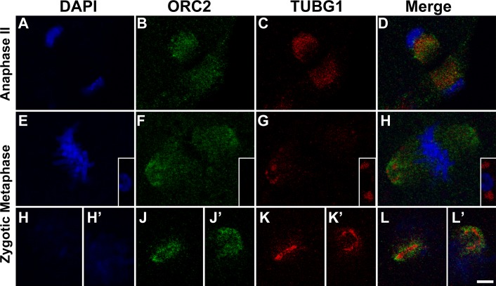
Colocalization of OCR2 and TUBG1 in anaphase II and zygotic mitosis. Zygotes were fixed and double immunostained for ORC2 (B, F, J, and J′) and for TUBG1 (C, G, K, and K′) and counterstained with DAPI (A, E, H, and H′). A–D) Anaphase II of the maternal chromosomes shortly after fertilization. ORC2 is localized to two foci between the separating chromosomes, as in Figure 1Bb (B). TUBG1 is closely associated with ORC2 but is closer to the DNA. E–H) ORC2 (F) and TUBG1 (G) are at the spindle poles. TUBG1 appears as a loose ring-like structure (G). The polar body demonstrates a more tightly focused TUBG1 signal, more similar to somatic cells (G, inset). H–L′) The two spindle poles from another zygote. In one spindle pole, TUBG1 appears as a linear signal (K) with ORC2 surrounding the TUBG1 (J), while in the second spindle pole, TUBG1 appears as a ring (K′) with ORC2 surrounding the TUBG1 ring (J′). Bar = 5 μm.

