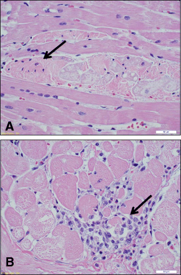
Figure 4.
Histopathology of left ventricular cardiac tissue from an affected pig. Sections from a 1 year-old affected male pig show myofibrillar degeneration (magnification = 30X). A. Fragmentation of myocytes, pyknotic nuclei (arrow) and loss of cross-striation. B. Aggregation of lymphocytes and loss of myofibrils (arrow).

