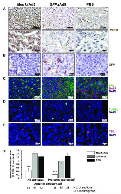
Figure 4. Assessment of proliferation, apoptosis and immune response in Men1+/− pituitary tumours at four-weeks after intra-tumoural injection of either Men1.rAd5, GFP.rAd5, or PBS (control).
A, Menin immunostaining, using DAB, and nuclei counterstaining with haematoxylin, at four-weeks. Menin expression was absent in Men1+/− pituitary tumours (PBS control and GFP.rAd5), but specific nuclear expression of menin (brown) was observed in Men1+/− pituitary tumours injected with Men1.rAd5 (p<0.04). Top and bottom panels represent views at x20 and x40 magnification respectively. B, GFP immunostaining, using DAB, and nuclei counterstaining with haematoxylin. Cytoplasmic GFP expression (brown, middle panel) was observed only in Men1+/− pituitary tumours that were injected with GFP.rAd5. C, Proliferation in prolactin expressing cells. Immunofluorescent labelling for prolactin (PRL, green) and BrdU (red) with nuclear DAPI counterstain (blue). BrdU labelling is reduced in Men1+/− pituitary tumours injected with Men1.rAd5. D, Apoptosis. Apoptotic cells (green) labelled using the TUNEL assay with DAPI counterstain (blue). Apoptosis was similar in the three groups. E, Immune response. Immunofluorescent labelling for CD3 (red) with DAPI counterstain (blue). Numbers of CD3 positive cells were similar in all three groups. F, Proliferation rates in Men1+/− pituitary tumours. Intra-tumoural injection of Men1.rAd5 resulted in a significant decrease in proliferation rates of prolactin-expressing cells and all pituitary tumour cells. Data shown as mean±SEM, ***p<0.000001 (Men1.rAd5 v GFP.rAd5 and Men1.rAd5 v PBS).

