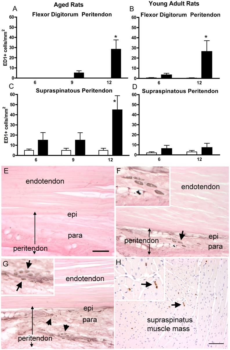
Figure 6. ED1-immunoreactive macrophages in flexor digitorum and supraspinatus peritendon regions of preferred reach limbs.
(A) ED1 cells in the flexor digitorum peritendon in aged rats, and (B) in young adult rats. (C) ED1 cells in supraspinatus endotendon of aged rats, and (D) in young adult rats. *p<0.05, compared to age-matched NC. (E) Photo of a flexor digitorum tendon of an aged-NC rat, and (F) in an aged 12-week HRLF rat. Arrow depicts ED1 cells in paratendon (ED1 is black; eosin counterstain), shown enlarged in the inset. (G) Photo of a distal cut end of a supraspinatus tendon of an aged 12-week HRLF rat. Arrows depict ED1 cells in paratendon, shown enlarged in inset. (H) Photo of the muscle mass of an aged 12-week HRLF rat showing ED1 cells. Arrow depicts ED1 cells (brown in color; eosin counterstain), shown enlarged in inset. Scale bar = 50 micrometers.

