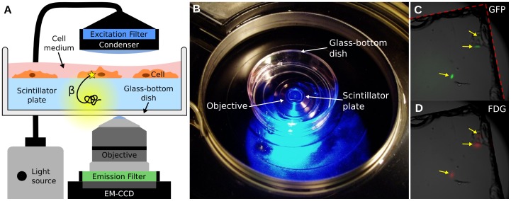
Figure 1. Overview of the radioluminescence microscope.
(A) Radioluminescence is produced within a scintillator plate following the emission of a beta particle from a radiotracer within a cell (yellow glow). The optical photons are captured by a high-numerical-aperture objective coupled to a deep-cooled EM-CCD camera. Emission and excitation filters used in combination with a light source allow for concurrent fluorescence and brightfield microscopy. (B) Photograph of the system showing a glass-bottom dish containing a scintillator plate immersed in cell culture medium and placed into the inverted microscope. (C) Three GFP-expressing HeLa cells located near the corner of a scintillator plate were localized using fluorescence microscopy (arrows). The edge of the scintillator plate is outlined in red. (D) After incubation with FDG (400 µCi, 1 h), these three cells also produced focal radioluminescence signal coincident with the fluorescent emission.

