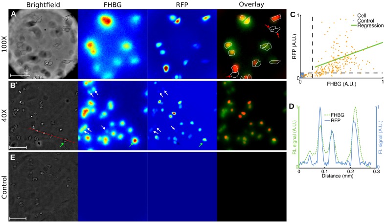
Figure 5. Radioluminescence imaging of gene expression in single cells.
Human cervical cancer cells (HeLa) transfected with a fusion PET/fluorescence reporter gene were incubated with FHBG (300 µCi, 2 h). (A) Brightfield (scale bar, 50 µm), radioluminescence (FHBG), and fluorescence (RFP) micrographs (objective, 100X/1.35 NA). Overlay shows FHBG radioluminescence (green), RFP fluorescence (red), and cell outline segmented from brightfield. Cells negative for RFP are also negative for FHBG (red arrows). (B) Same as (A), but with a 40X/1.3 NA objective (scale bar, 100 µm). White arrows indicate cells with weak fluorescence intensity but substantial radioluminescence intensity. The green arrow points to a cell with no RFP expression but ambiguous radioluminescence intensity. (C) Scatter plot of FHBG vs. RFP uptake, computed for 245 cells (light red dots) and 100 control ROIs (blue dots). Arbitrary units. (D) Radioluminescence and fluorescence shown along a line profile [red dashed line in (A)]. (E) Same experiment as (A,B), but using control wild-type HeLa cells (scale bar, 100 µm).

