
Figure 2.
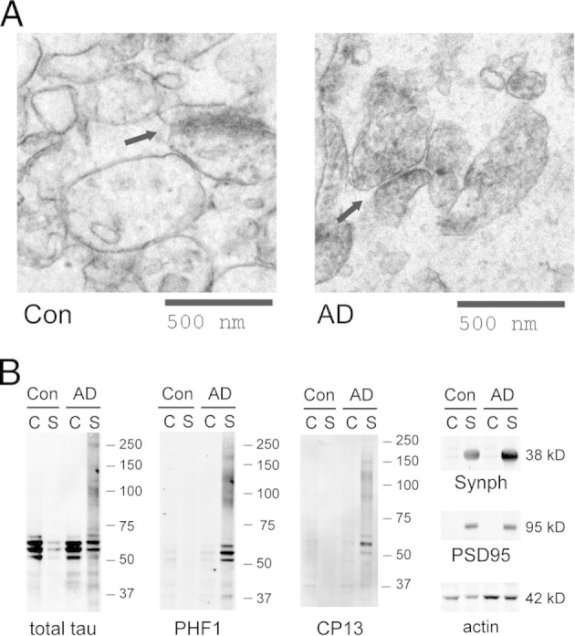
A: Synaptoneurosomes from human cortices examined under electron microscopy show well-preserved synaptic structures (arrows). No protein fibrils were observed in control or AD samples. B: In AD-affected brains, p-tau oligomers (detected using PHF1 and CP13) accumulate in the synaptoneurosome (S) but not in the cytosol (C). Synaptophysin (Synph) and PSD95 serve as synaptic markers. Images are representative of three independent experiments.