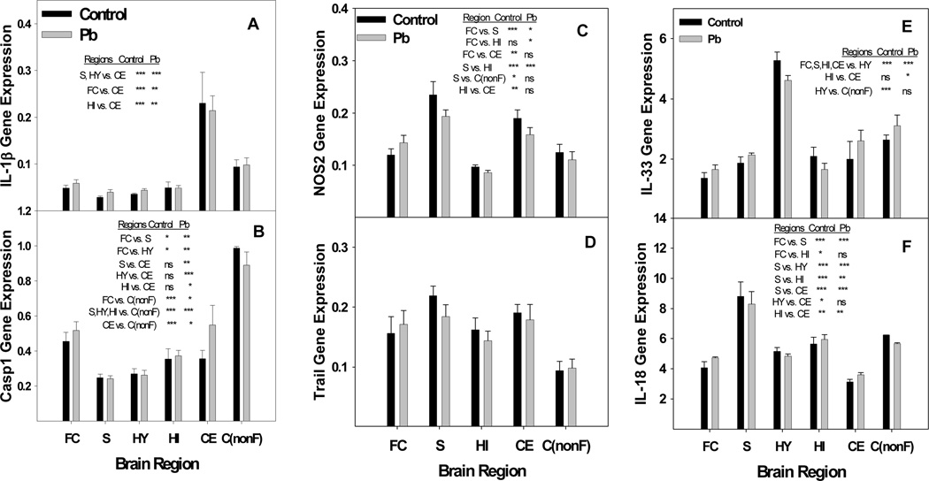
Fig. 9.
Developmental Pb exposure perturbs regulated gene expression between brain regions of select genes associated with inflammation. A) IL-1β, B) Caspase 1, C) NOS2, D) Trail, E) IL-33, and F) IL-18. Brain region total RNA was isolated as described in the methods from female mice exposed to 0.1 mM Pb from gd8 to pnd21 and distilled water controls. NOS2 data for striatum and hippocampus include an N of 3 litters and all non-frontal cortex data represents an N of 2 litters each for control and Pb-exposed. All other data represent an N of 4 litters for control and Pb-exposed mice. Statistics were performed by ANOVA analysis with Bonferroni post-test restrictions. Asterisks indicate the following P values: * p<0.05, ** p<0.01, *** p<0.001. No significant difference is indicated by ns. Regions are Frontal Cortex (FC), Striatum (S), Hypothalamus (HY), Hippocampus (HI), and Cerebellum (CE), non-frontal Cortex C(nonF). Data are presented as mean ±S.E.M.

