Fig. 1.
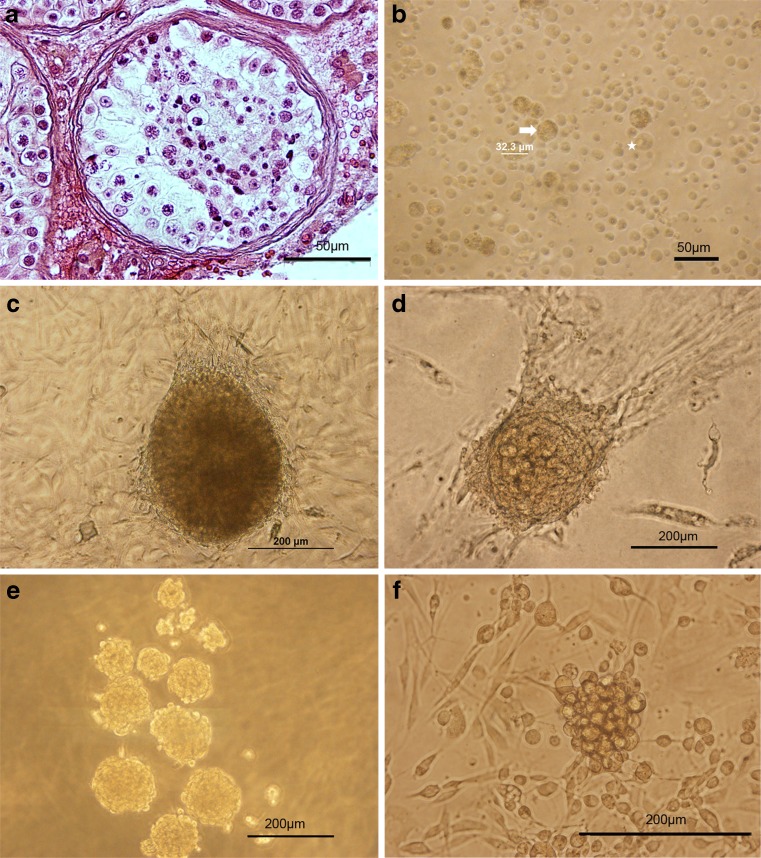
Isolation and culture of SSCs from testicular tissues of non-obstructive azoospermic (NOA) patients. a Histological appearance of the testes biopsy obtained from azoospermic patient with complete maturation arrest that was stained with H& E and showed spermatogenic cells in tubules. b Cell population obtained from the seminiferous tubules after two steps enzymatic digestion contained different cell types, sizes and morphology. Spermatogonia could be identified as round cells with a large nucleus, one nucleoli and cytoplasmic inclusions (asterisk); whereas, Sertoli cells were large cells with a granular appearance (arrows). After overnight incubation, the non-adhering cells were collected and cultured. c–f The morphology of clusters growing on top of monolayer of somatic cells shows three types of colonies or clusters as observed during 2 months of cultivation. First, ES-like colonies were sharply edged and compact (c); while, big (d) and small (e, f) GSC clusters were smaller and clumpy and their cells were individually recognizable. We assayed the GSC clusters. Scale bars: A, B = 50 μm and C-F = 200 μm

