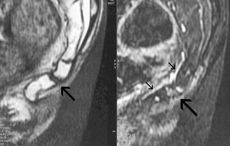
Fig. 1.
A 50-year-old man presenting hypermobility of the coccyx (30° of flexion in the seated position). Left: T1-weighted image showing the coccygeal anatomy. Right: T2-weighted hypersignal (with a short tau inversion recovery sequence) of the intercoccygeal joint and the adjacent vertebral end plates (image on the right, large arrow). Note the presence of a large drainage vein (image on the right, small arrows) on the ventral aspect of the coccyx

