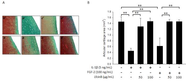
Figure 6. LfcinB inhibits IL-1β/FGF-2-mediated proteoglycan depletion in articular cartilage ex vivo.

(A) Full-thickness cartilage explants with 4 mm diameters in serum-free media (plus mini-ITS™+ Premix) were treated with FGF-2 (100 ng/mL) or IL-1β (5 ng/mL), in the presence or absence of LfcinB (50 and 100 μg/mL), for 11 days. The explants were fixed in 4% paraformaldehyde and embedded in paraffin. Safranin O Fast Green staining was adopted to assess gross proteoglycan content in the extracellular matrix. (a & a’) Control; (b) FGF-2 (100 ng/mL); (c) FGF-2 plus LfcinB (50 μg/mL); (d) FGF-2 plus LfcinB (100 μg/mL); (b’) IL-1β (5 ng/mL); (c’) IL-1β plus LfcinB (50 μg/mL); (d’) IL-1β plus LfcinB (100 μg/mL). (B) Stained sections were analyzed for proteoglycan staining-positive areas using OsteoMeasure software. Superficial zone and middle zone were included into analyses, and cartilage areas were calculated based on sections from 3 different donors. **p<0.01.
