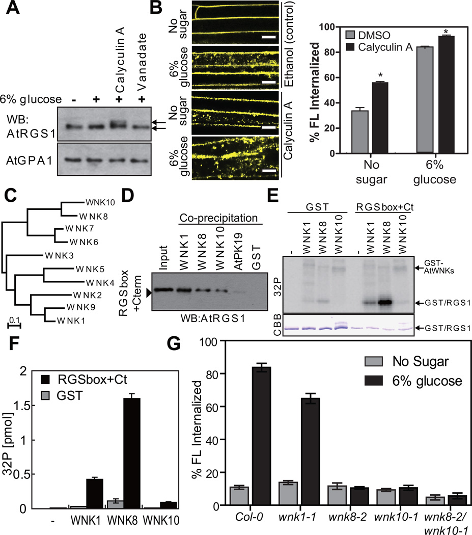
Figure 3. In vivo and In vitro function of AtWNK8.
(A) In vivo phosphorylation of AtRGS1. Seedlings expressing AtRGS1-TAP were pretreated with 100 nM calyculin A and 10 mM sodium orthovanadate for 3 h followed by 6% D-glucose stimulation for 90 min. AtRGS1-TAP or AtGPA1 in seedling lysates was separated on a 12.5% Anderson’s gel and detected by immunoblot with peroxidase anti-peroxidase or anti-AtGPA1 antibody. (B) Four-day-old AtRGS1-YFP expressing seedlings were treated with phosphatase inhibitors, calyculin A, for 2 h followed by 6% glucose treatment or not (No glucose) for 1 h prior to imaging epidermal cells. Scale bars = 10 µm. Error = SEM, n = 5. (C) Phylogenetic tree of the AtWNK-family kinases. Full-length amino acid sequences were aligned with CLUSTAL W implemented in CLC Genomics Workbench using the following settings; Gap open penalty, 10; Gap extension penalty 1. The neighbor joining tree (1000 bootstrap replicate) was created with the aligned sequences. (D) In vitro binding between AtRGS1 and AtWNKs. Recombinant RGSbox+Cterm was tested for interaction with GST (negative control) or GST-AtWNKs using glutathione-Sepharose, and detected by immunoblot analysis using an anti-AtRGS1 antibody. (E) In vitro phosphorylation of AtRGS1 by AtWNK kinases. Recombinant GST or His-RGSbox+Cterm was incubated with GST-AtWNKs in reaction buffer containing γ32P-ATP. Proteins were separated on SDS-PAGE. (F) Radioactivity incorporated into the GST/RGS1 bands. Phosphorylation levels of three independent experiments were quantified in (E). Error bars = SEM. (G) Quantitation of sugar-induced AtRGS1 internalization in AtWNK-null mutants. Seedlings of Col-0, wnk1-1, wnk8-1, wnk8-2 or wnk10-2 transiently expressing AtRGS1-YFP were treated with 6% D-glucose for 30 min. WNK# denotes AtWNK members in panels C-F. Error bars = SEM, n = 5. Quantitation of fluorescence is described in Methods.

