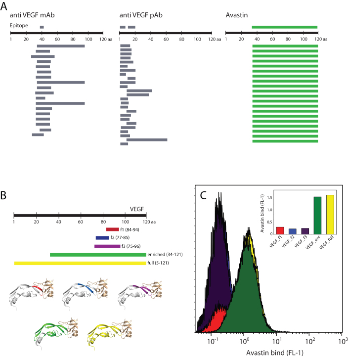
Figure 5. S. carnosus displays folded VEGF which is recognized by Avastin.
(A) Epitope mapping of anti-VEGF antibodies using S. carnosus surface display of VEGF fragments. Commercial mAbs and pAbs bind to small VEGF fragments, allowing for fine epitope mapping. In the Avastin assay, a single, large VEGF fragment is enriched, indicating a conformational epitope. (B) VEGF fragments containing potential Avastin epitopes. The fragments are shown colored on the VEGF:Avastin Fab co-crystal structure (PDB 1BJ1), which shows a conformational epitope. (C) Avastin binding to S. carnosus clones of VEGF fragments (colored as in (B)). Avastin does not bind to smaller epitope fragments but requires large and presumably folded VEGF fragments.

