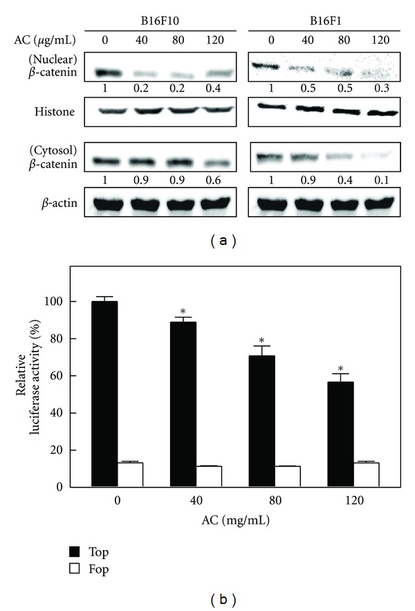
Figure 3.

AC inhibited β-catenin nuclear translocation and transcriptional activation in melanoma cells. (a) B16F10 and B16F1 cells were treated with or without AC (40–120 μg/mL) for 24 h. The levels of β-catenin in the nuclear and cytoplasmic fraction were determined by Western blot. Histone H3 and β-actin were used as an internal loading control for nuclear and cytoplasmic fractions, respectively. The photomicrographs shown in this figure are from one representative experiment performed in triplicate, with similar results. (b) B16F10 cells were transiently transfected with TOPFlash or FOPFlash plasmids by using lipofectamine and then incubated with or without AC (40–120 μg/mL) for 24 h. Cell lysates were mixed with luciferace reagents and quantified by luminometer. Relative β-catenin activity was calculated by dividing the relative luciferase unit (RLU) of treated cells by the RLU of untreated cells. The results are presented as the mean ± S.D of three independent assays. *Significant difference in the control versus sample group (P < 0.05).
