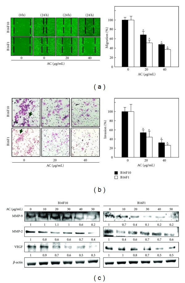
Figure 6.

AC inhibits the invasion and migration of melanoma cells. (a) AC-induced inhibition of melanoma migration was measured by in vitro wound healing assay as described in Section 2. Cells were seeded in a 24-well plate, and mechanically scratched to make a wounded area in the culture cells. After incubation with AC, migration was observed using a phase-contrast microscope (100x magnification) at 0 and 24 h, and the closure of the area was calculated. (b) Melanoma invasion was monitored by Transwell chamber assay. Cells were pretreated with AC, and, after 24 h, cells invading under the membrane were photographed (200x magnification). The inhibition percentage of invading cells was quantified and expressed on the basis that untreated cells (control) represented 100%. The results are presented as the mean ± S.D of three independent assays. *Significant difference in comparison to the control versus sample group (P < 0.05). (c) B16F10- and B16F1-mediated down-regulation of MMP-9, MMP-2, and VEGF expression was determined by Western blot analyses. Cells were treated with AC for 24 h. Equal amounts of protein samples (50 μg) were resolved by 8–15% SDS-PAGE. The photomicrographs shown here are from one representative experiment repeated three times with similar results.
