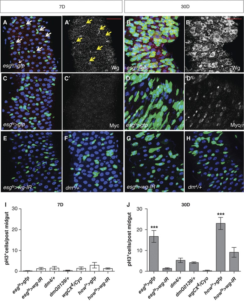
Figure 8.
Wg and Myc are upregulated in ageing Drosophila midguts. (A–D′) Midguts expressing gfp under the control of the esg-gal4 driver stained with anti-Wg (A–B′) or anti-Myc (C–D′) at 7 and 30 days after adult eclosion. Arrows in (A, A′) point to a subset of esg+ve cells that also express Wg. Note the increase in esg+ve cells and upregulation of Wg and Myc in aged guts. (E–H) Posterior midguts from 7- or 30-day-old adults, expressing RNAi for wg under the control of the esg-gal4 driver (E, G) and midguts from age-matched flies heterozygous for a loss of function allele of myc (F, H). (I, J) Quantification of pH3+ve cells in the genotypes indicated at 7 (I) or 30 (J) days (***P<0.0001 one-way ANOVA with Bonferroni’s multiple comparison test). Partial loss of wg or myc suppressed the age-dependent ISC proliferation hyperproliferation. Scale bars: 20 μm.

