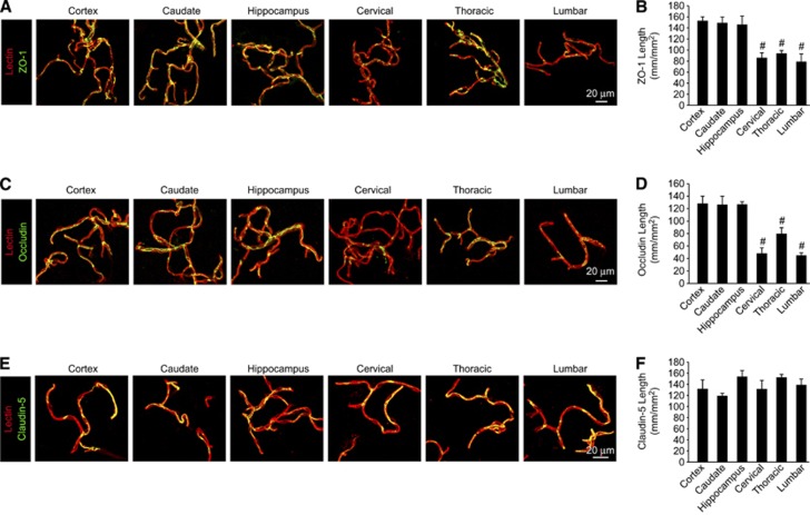
Figure 4.
Reduced pericyte coverage along blood–spinal cord barrier is associated with reduced tight junction protein expression. (A, C, E) Confocal microscopy analysis of tight junction zonula occludens-1 (ZO-1) (A), occludin (C), or claudin-5 (E) immunodetection (green) in acutely isolated lectin-positive capillaries (red) from 2-month-old mouse cortex, caudate, hippocampus, cervical, thoracic, and lumbar spinal cord. (B, D, F) Quantification of endothelial ZO-1 (B), occludin (D), or claudin-5 (F) tight junctional length in acutely isolated vessel preparations from the brain and spinal cord regions. Mean±s.e.m. n=3 to 4 preparations per group (five mice per preparation); #P<0.05 when compared with the brain regions.

