Figure 1.

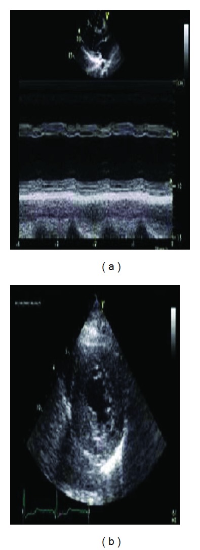
(a) Parasternal long-axis view showing the left ventricular dilatation and advanced global hypokinesis. (b) Parasternal short axis view showing the septal and posterior wall hypertrophy.

(a) Parasternal long-axis view showing the left ventricular dilatation and advanced global hypokinesis. (b) Parasternal short axis view showing the septal and posterior wall hypertrophy.