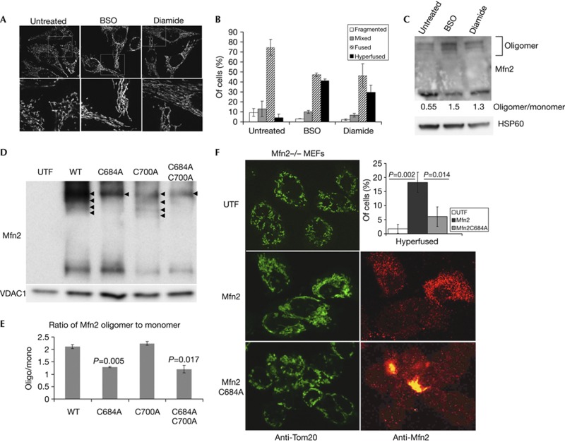
Figure 4.
GSSG-induced Mfn2 oligomers through Cysteine 684 are required for fusion in vivo. (A) Treatment of HeLa cells with BSO (100 μM for 24 h) or Diamide (100 μM for 1 h), leads to increased fusion in vivo, as revealed by Tom20 staining. (B) Quantification of at least 30 cells from triplicate reactions treated as in A, error bars show mean+s.d. (C) Western blots and quantification of the increase in oligomeric Mfn2 on inhibition of glutathione synthesis. (D) Wild-type Mfn2, Mfn2C684A and/or Mfn2C700A constructs were transfected into Mfn2−/− MEFs as indicated. Mitochondria were isolated, treated with 0.5 mM GSSG and Mfn2 oligomers observed by western analysis following non-reducing electrophoresis. VDAC1 was probed as a load control. Arrowheads indicate the presence of oligomeric forms of Mfn2. (E) The ratio of oligomer:monomer was quantified from two blots and the P-value versus the wild type is indicated. (F) Confocal analysis of mitochondrial morphology within Mfn2−/− MEFs either untransfected (UTF), or transfected with Mfn2 or Mfn2C684A. Mitochondria are labelled with Tom20 (green), and transfected cells are confirmed using monoclonal anti-Mfn2 antibodies (red). The percentage of transfected cells showing hyperfused mitochondria in each condition is shown in the top right panel. Data were obtained from two experiments counting at least 50 cells for each condition; P-values were determined by Student’s T-test, error bars show mean+s.d. BSO, L-buthionine-sulfoximine; GSSG, oxidized glutathione; MEF, mouse embryonic fibroblast; Mfn2, Mitofusin 2; WT, wild-type.

