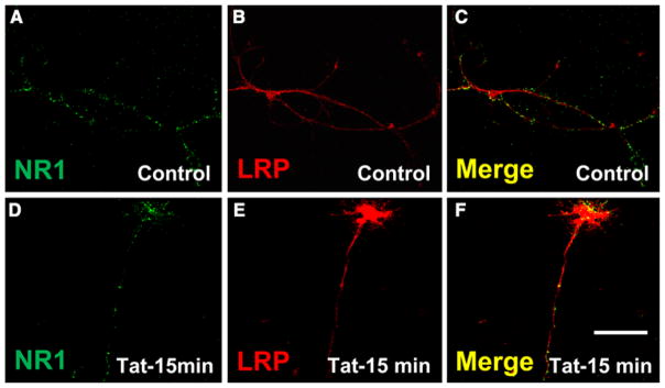
Fig. 5.
Rat hippocampal neurons express LRP/NMDAR complexes, but the distribution of these proteins is not altered by tat treatment. Confocal microscopy indicates that tat treatment (100 ng/ml, 15 min) did not alter the colocalization of LRP and NR1 on the surface of rat hippocampal neurons. Double immunofluorescence labeling for LRP (Cy3 staining, red) and NR1 (FITC staining, green) shows LRP and NR1 in control (a–c) and tat (d–f) treated cultures. Specificity was confirmed by replacing the primary antibody with a non-specific myeloma protein of the same isotype or the appropriate polyclonal reagent (data not shown). Bar: 20 μm. (n = 3)

