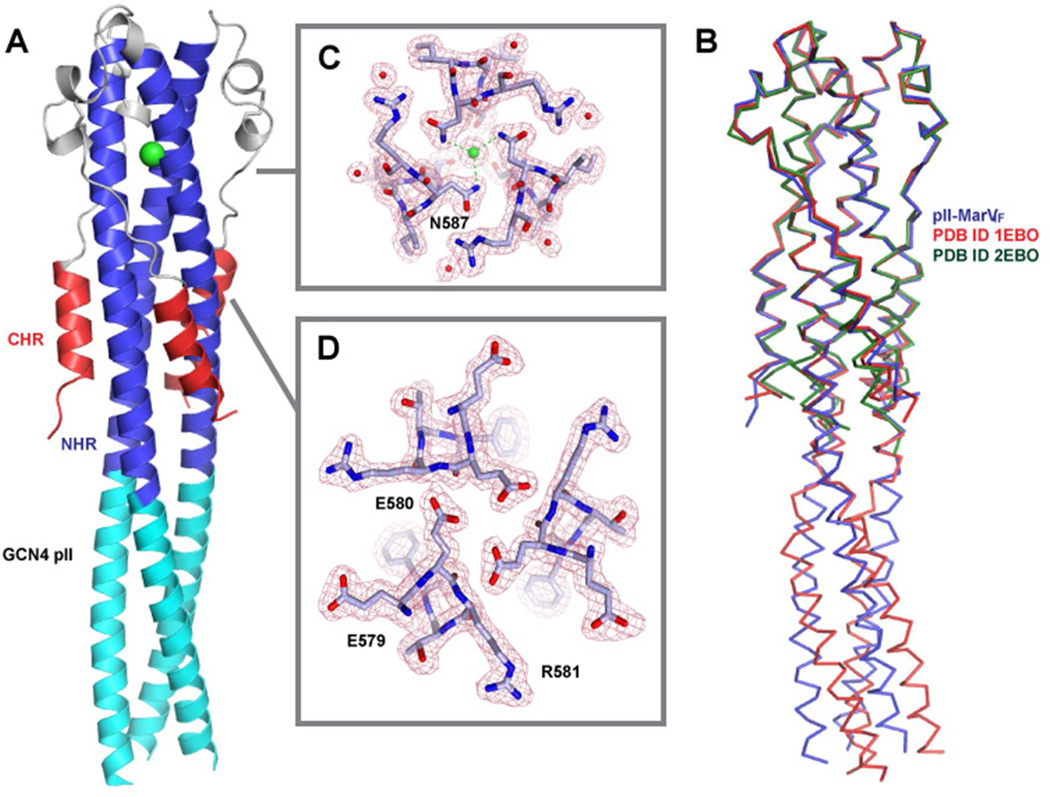
Figure 3. Structural Features of the pII-MarVIF Crystal Structure.
(A) Overall depiction of the fold, with secondary structural elements colored according to the cylinders in Figure 1. The chloride ion bound by N587 is shown as a green sphere and depicted with electron density in panel C. (B) Overlay of pII-MarVIF with the two reported EBOV GP2 structures (PDB ID 1EBO, Weissehorn et al., ref. 20; and PDB ID 2EBO, Malashkevich et al., ref. 21). The structural alignment was performed with the MARV/EBOV GP2 segments only. (C&D) View down the NHR trimer axis and electron density for regions near the chloride-binding site (C) and the E579 residue oriented toward the core (D).

