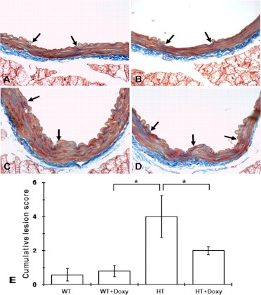
Fig. 2.
Spontaneously developed lesions in aortas of 9-month-old mice. A to D, typical representations of spontaneous lesions. Sections were stained with Masson's trichrome. A, lesion grade 2 in WT mouse. B, section of aorta from lesion grade 2 in HT mouse. C, lesion grade 3 lesion; a large defect in the internal elastic lamina and significant subintimal spindle cell proliferation with deposition of collagen. D, lesion grade 3, similar to grade 4 but with a defect area much larger. E, cumulative lesion score (lesions ≥ grade 2) in each group. Values are means ± S.E.M. *, p < 0.05, ANOVA with Tukey post hoc test.

