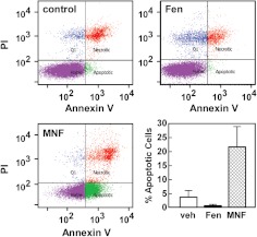
Fig. 5.
MNF induces apoptosis in HepG2 cells. Serum-depleted HepG2 cells were treated with vehicle (top left), Fen (1 μM) (top right), or MNF (1 μM) (bottom left) for 24 h, stained with Annexin V and PI, and then analyzed by flow cytometry. Representative profiles are shown. The fraction of annexin V-positive HepG2 cells that were apoptotic was quantitated and represents results from two independent experiments, each performed in duplicate dishes (lower right). Data are expressed as means ± S.E. (n = 4).

