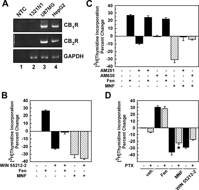
Fig. 6.
Role of CBR activation in the antiproliferative action of MNF in HepG2 cells. A, total RNA was extracted from HepG2, 1321N1, and U87MG cells, and then analyzed semiquantitatively by PCR. A nontemplate control (NTC) has been included (lane 1). GAPDH, glyceraldehyde-3-phosphate dehydrogenase. B and C, serum-depleted HepG2 cells were incubated with the CBR agonist WIN 55,212-2 (Win; 1 μM) (B) or antagonists AM251 (1 μM) or AM630 (0.5 μM) (C) for 1 h followed by the addition of vehicle, Fen (0.5 μM), or MNF (0.25 μM) for 24 h. D, serum-depleted HepG2 cells were pretreated without or with pertussis toxin (PTX; 50 ng/ml) for 16 h followed by the addition of vehicle, Fen (0.5 μM), MNF (0.5 μM), or WIN 55,212-2 (0.5 μM) for 24 h. In B to D, the levels of [3H]thymidine incorporation were measured. Quantification of percentage change in [3H]thymidine incorporation versus control is expressed as means ± S.D. and represents results from three (B and C) or two (D) independent experiments, each performed in triplicate dishes. *, p < 0.05.

