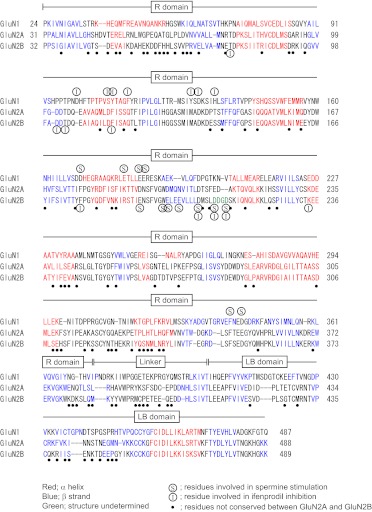
Fig. 3.
Amino acid sequences of the R domains and immediately distal regions in GluN1, GluN2A, and GluN2B. Secondary structure alignment was performed as described under Materials and Methods. The positions of α helices and β strands are shown in red and blue, respectively. Amino acid residues shown in (S) and (I) are residues that influence spermine stimulation and ifenprodil inhibition, respectively. Amino acid residues shown in ● are not conserved between GluN2A and GluN2B.

