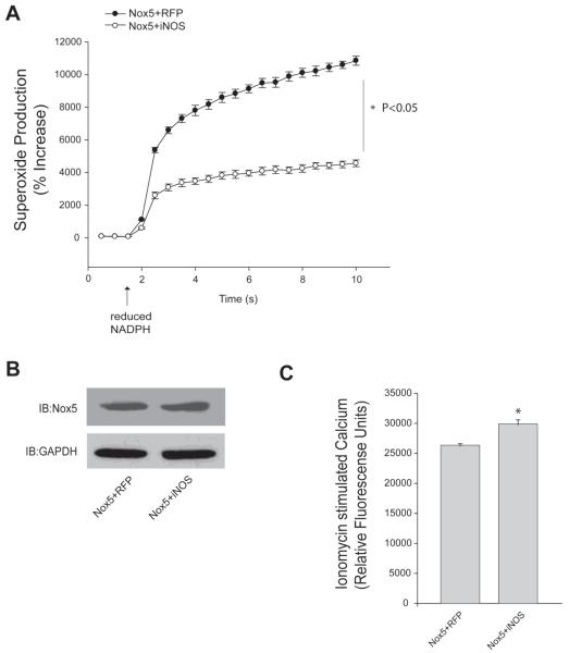
Figure 5. NO reduces Nox5 enzyme activity in a cell-free activity assay but does not reduce intracellular calcium levels.
(A) COS-7 were co-transfected with Nox5 and either iNOS or RFP (control). Cells were lysed and cell-free activity of Nox5 was measured in the presence of CaCl2 (1mM), NADPH (200μM) and FAD (100μM). Superoxide levels were determined via L-012 chemiluminescence and expressed as % increase above unstimulated levels (means ± S.E., n =6 * p<0.05 versus RFP). (B) Expression of Nox5 was determined by Western blot with anti-HA and GAPDH was used as loading control. (C) Intracellular Ca2+ was measured after addition of ionomycin (1μM) in transfected COS-7 cells using the Fluo-4. Results are presented as mean ± S.E. (n = 8 * p<0.05 versus RFP).

