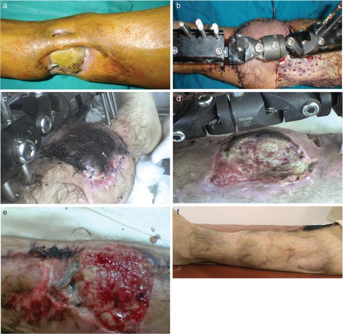
Fig. 4.
Pre-operative clinical view (a) of a non-healing distal lower extremity wound with tibia necrosis for the patient in Case No. 19. Intra-operative view showing the harvested peroneal based perforator flap with dimensions of 17 cm in length and 12 cm of width (b). Post-operative view showing the superficial necrosis of the distal third of the flap after venous congestion (c). The flap was revised and surgically debrided (d) with adequate granulation tissue 7 days after the revisional surgery (e) when skin grafting was performed. Post-operative clinical outcome at 6 months follow-up (f).

