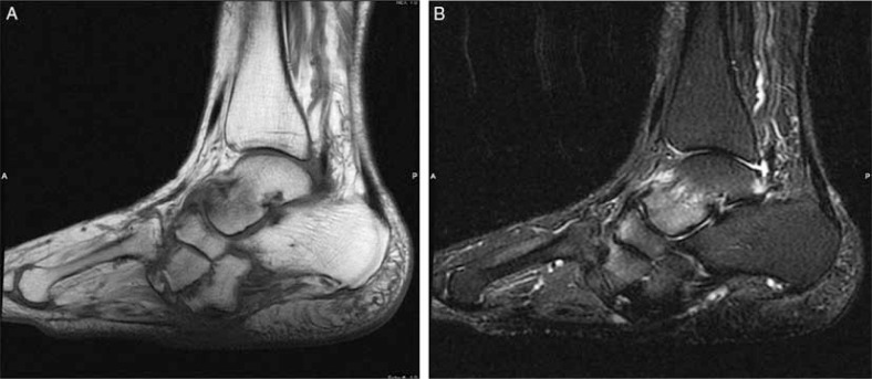
Fig. 1.
Skin callus and talar plantar flexion deformity in a diabetic patient with neuroarthropathy. Sagittal T1 (A), and T2-weighted with fat-suppressed (B) images revealed focal hypointense area in subcutaneous fat in the midfoot in both sequences (arrows) with no accompanying soft tissue changes consistent with callus. Subchondral marrow edema at intertarsal joints is a result of neuroarthropathy.

