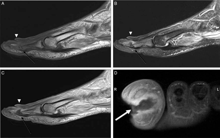
Fig. 3.
Cellulitis, fistula and associated osteomyelitis and septic arthritis of the first toe (A–D). Sagittal T1 (A), T2-weighted fat-suppressed (B), and post-contrast T1-weighted (C), and short-axis T1-weighted fat-suppressed (D) images demonstrate a high T2 signal and significant skin enhancement after administration of contrast demonstrating cellulitis (arrowheads). There is a deep ulcer in the medial portion of the first toe and fistula (white arrow) traversing the distal phalanx. Sagittal images also demonstrated the abnormal signal of the proximal and distal phalanges due to osteomyelitis and accompanying septic arthritis (black arrow).

