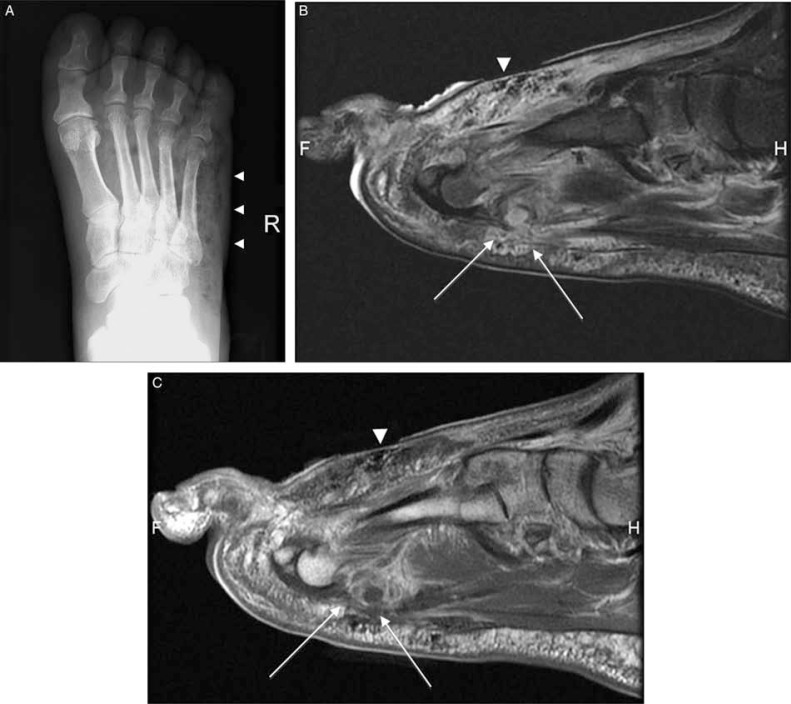
Fig. 4.
Gas gangrene and pyomyositis in a patient with diabetes (A–C). Plain radiography (A), and sagittal T2-weighted fat-suppressed (B) demonstrate extensive soft tissue gas (arrowhead) in the dorsum of the foot. In T2-weighted image, diffuse signal increase in plantar muscles and fluid like focal areas (arrows) that demonstrated peripheral rim enhancement (arrows) in post-contrast T1-weighted images (C) consistent with pyomyositis.

