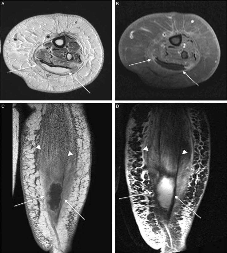
Fig. 6.
Diabetic myonecrosis of the left posterior calf muscles in a poorly controlled diabetes patient in an extremely painful and tender left leg. Transverse fat-saturated T2–weighted (A), post-contrast, transverse fat-suppressed T1 (B), and coronal T1-weighted (C), and T2-weighted-fat suppressed (D) images shows a cystic non-enhancing area within gastrocnemius muscle (arrows) and swollen edematous posterior calf muscles (arrowheads) consistent with myonecrosis. Note the extensive cellulitis and superficial fascial inflammation but no signs of soft tissue ulcer or sinus tract.

