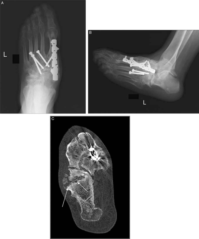
Fig. 9.
Midfoot reconstruction in a patient with unstable neuropathic osteoarthropathy (A–C). Anteroposterior (A), and lateral (B) plain radiographs demonstrated neuropathic changes in the midfoot region and complex realignment and fusion (A). Joint destruction, osseous fragmentation and subchondral cyst formation (arrows) better delineated compared to plain radiographs in transverse CT image (C).

