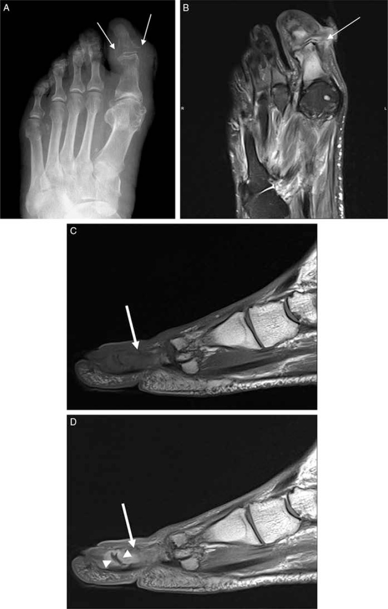
Fig. 12.
Septic arthritis and associated osteomyelitis in a diabetic patient. Plain radiograph (A) demonstrates focal soft tissue swelling, demineralization (arrows) in periarticular region in distal interphalangial joint of the first toe. Long-axis T2-weighted fat-suppressed image (B) demonstrated an ulcer and sinus tract (thin white arrow) extending to the joint space. There is a synovial enhancement (arrowhead) and abnormal intramedullary signal (thick white arrow) that is extending from the joint surface in pre-(C) and post-contrast T1-weighted (D) images consistent with septic arthritis and accompanying osteomyelitis.

