Fig. 4.
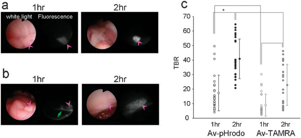
In vivo fluorescence endoscopic images in peritoneal tumor bearing mice with Av-pHrodo (a) or Av-TAMRA (b). The pink arrow heads show the tumor nodules. The tumors were visualized with both Av-pHrodo and Av-TAMRA 1 h after the injection, but fluorescence from excess injectate in the peritoneal cavity (green arrow) was also detected. Two hours after the imaging probe injection, the tumor nodules were clearly visualized by both Av-pHrodo and Av-TAMRA, although the background signal was lower for Av-pHrodo. The measured tumor-to-background ratios (TBRs) are summarized in (c). The highest TBR was accomplished by Av-pHrodo 2 h after the injection. (*p < 0.001, Mann-Whitney U test).

