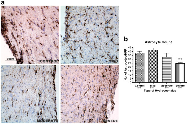
Figure 6.
Astrocytic activation in hydrocephalus. a: GFAP immunoreactive cells in the sub-ependymal layer showing astrocytic activation (arrows) in the hydrocephalic rats , particularly in the mild group. b: Astrocyte count in the same region. Astrocyte count was raised in mild hydrocephalus (not significant) but gradually decreased as ventriculomegaly progressed and was significantly reduced in severe hydrocephalus (*** p<0.001 vs control). Data are means ± SEM. (HPF = High power field). Scale bar 75 μm).

