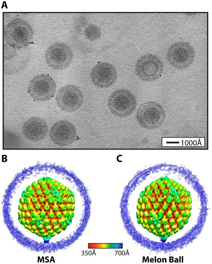
Figure 1. Visualization of a distinctive tegument structure associated with the portal vertex in HSV-1 virions.
(A) Projection image through a tomogram of HSV-1 virions embedded in vitreous ice (Video S2). (B–C) The symmetry-free virion averages generated from 213 subtomograms using MSA-guided classification and melon ball alignment methods, respectively. The maps are shown radially colored, with non-capsid densities trimmed to reveal the underlying capsid. Views are shown looking down a 2-fold axis of symmetry with the portal vertex at the bottom.

