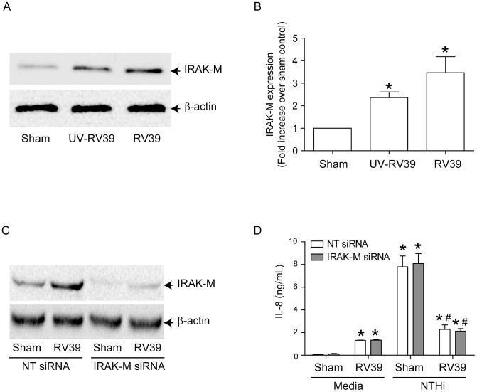
Figure 5. IRAK-M is not responsible for RV-induced suppression of NTHi-stimulated IL-8 in airway epithelial cells.
(A and B) BEAS-2B cells were infected with sham, RV39 or UV-RV39 as described under Figure 2. Aliquots of total cell lysates corresponding to equal amount of protein from were subjected to Western blot analysis with IRAK-M antibody. (A) Representative blot showing IRAK-M expression. (B) Band intensities were quantified by NIH image and levels of IRAK-M was normalized to β-actin and expressed as fold increase over sham infected cells. (C) BEAS-2B cells were transfected with non-targeting (NT)- or IRAK-M siRNA and knockdown of IRAK-M was confirmed by Western blot analysis. (D) Similarly transfected cells were infected with sham or RV followed by NTHi as described in Figure 3 and IL-8 determined in the media. Data represents mean±SEM calculated from 3 independent experiments performed in triplicate (* p≤0.05, ANOVA, different from sham; #p≤0.05, ANOVA, different from sham/NTHi).

