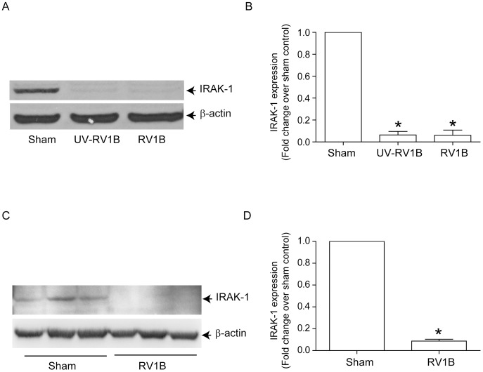
Figure 10. RV infection decreases IRAK-1 expression in alveolar macrophages in vitro and in mice lungs in vivo.
(A and B) MH-S cells were infected with sham, RV1B or UV-RV1B as described in figure 2 and incubated for 24 h. Cells were lysed in RIPA buffer and aliquots of cell lysates containing equal amounts of protein was subjected to Western blot analysis with IRAK-1 antibody. (C and D) Mice were infected with sham or RV1B, sacrificed two days later and lung lysates subjected to Western blot analysis to determine IRAK-1 expression. (A and C) Image presented is a representative of three independent experiments and 3 different animals respectively. (B and D) Band intensities of IRAK-1 were normalized to β-actin and expressed as fold increase over sham control. Data represents mean and SEM calculated from 3 independent experiments in B and 4 animals in D (* p≤0.05, ANOVA, different from sham).

