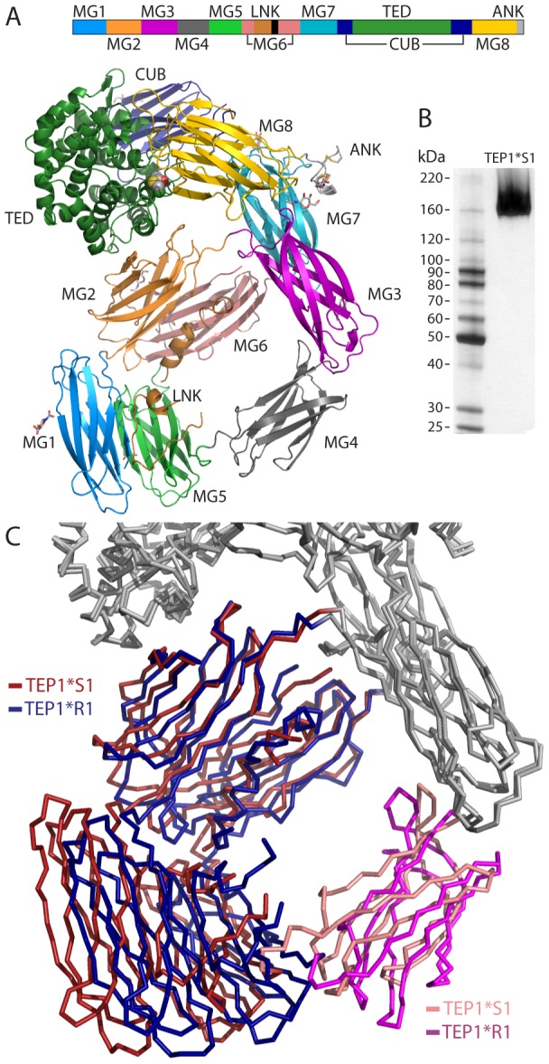
Figure 1. Overview of TEP1*S1 structure.

(A) Sequence schematic and three-dimensional structure of TEP1*S1. The mature protein commences with domain MG1 (mid-blue) followed by MG2 (orange), MG3 (purple), MG4 (mid-grey), MG5 (light green), MG6 (pink), LNK (light brown), MG7 (light blue), CUB (navy blue), TED (dark green), MG8 (yellow) and ANK (light grey). (B) Silver-stained SDS-PAGE of redissolved crystals confirms structure corresponds to full-length TEP1*S1. (C) Rigid body domain motions between TEP1*S1 and TEP1*R1. The MG1–2 and MG5–6 domains (*S1 red, *R1 blue) rotate by 11° relative to the remainder of the protein, with the MG4 domain (*S1 pink, *R1 magenta) acting as a hinge.
