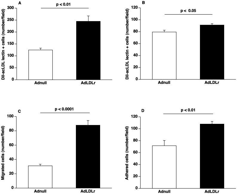
Figure 1. Lipid lowering gene transfer increases EPC number and enhances EPC function.
(A) Bar graph showing the number of Dil-acLDL FITC-isolectin double positive cells after 7 days of ex vivo culture of spleen mononuclear cells isolated at day 14 after Adnull transfer or AdLDLr transfer in C57BL/6 LDLr−/− mice (n = 5 for each group). (B) Bar graph illustrating the number of Dil-acLDL FITC-isolectin double positive cells after 7 days of ex vivo culture of bone marrow mononuclear cells isolated at day 14 after Adnull transfer or AdLDLr transfer in C57BL/6 LDLr −/− mice (n = 6 for each group). (C) Bar graph showing the number of migrated EPCs in modified Boyden chambers. After 7 days of culture, spleen EPCs isolated at day 14 after transfer from Adnull mice or AdLDLr treated C57BL/6 LDLr−/− mice were seeded in the upper chamber. The number of migrated cells per microscopy field was quantified after 5 hours (n = 12 for each group). (D) Bar graph illustrating the number of EPCs adhered to fibronectin-coated plates. After 7 days of culture, spleen EPCs isolated at day 14 after transfer from Adnull injected mice (n = 6) or AdLDLr treated mice (n = 6) were allowed to adhere onto fibronectin-coated plates for 30 minutes. Following vigorously washing with PBS, the number of adherent cells was counted under the microscope. Data are expressed as means ± SEM.

