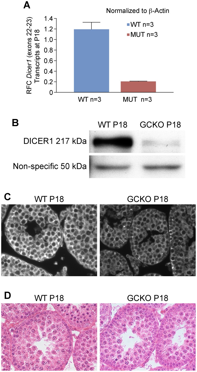
Figure 3. Levels of Dicer1 transcripts and DICER1 protein in GCKO testis samples are significantly reduced by P18.
(A) In comparison to WT testes, real-time qRT-PCR shows a 6-fold reduction in Dicer1 RNase III endonuclease transcripts in GCKO testes by P18 (*P = 0.018). (B) Western blot analysis using rabbit antibody against the N-terminal helicase domain of DICER1 and HRP-conjugated goat anti-rabbit IgG antibody shows reduced protein expression in GCKO testes by P18. The arrows point to the 217 kDa DICER1 protein and to the equivalent loading of 50 kDa protein from WT and GCKO testes. (C) Immunofluorescence detection of DICER1 protein in WT and GCKO testes at P18 using the same antibody used for western blotting. In WT sections, DICER1 localizes to the cytoplasm of most cell types populating the P18 testis, including Sertoli cells, spermatogonia and spermatocytes. At this developmental time point, secondary spermatocytes and spermatids are not present. In the P18 GCKO testis sections, DICER1 appears primarily in the cytoplasm of Sertoli cells located near the basement membrane with scant detection of DICER1 protein in the cytoplasm of spermatogonia and pachytene spermatocytes (magnification = 40x). (D) Cross sections of WT and GCKO testes on P18 stained with HE show no major differences in cellular composition suggesting that the changes in in Dicer1 transcript and protein levels in the GCKO testes were not explained by differences in cell populations.

