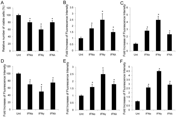
Figure 2. Impact of IFN-mediated β-catenin regulation on proliferation and apoptosis in HCC.
HepG2 (A) and Huh7 (D) cells were left untreated or treated with IFNα (100 ng/ml), IFNγ (100 ng/ml) or IFNλ (100 ng/ml) for 72 hrs. Cell viability was determined at 72 hrs. The abscissa represents the types of stimulation. The ordinate represents percentage of live cells relative to the mock treated cells. Apoptosis in HepG2 (B, C) and Huh7 (E, F) was measured by TUNEL assay and flow cytometry targeting active caspase 3. The ordinate represents fold increase of fluorescence intensity relative to the untreated cells. Data represent a minimum of three experiments and asterisks denote p<0.05 in comparison to untreated samples.

