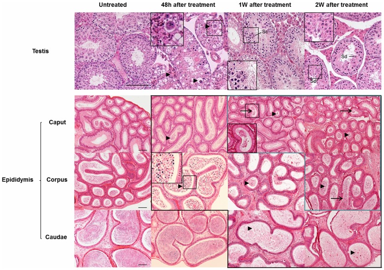
Figure 1. Morphology of normal and heat treated testes was revealed by hematoxylin and eosin staining.
Microscopic examination reveals that degenerated and apoptotic germ cells (black triangles) are visible in testes harvested 48 hours after a 42°C heat shock treatment, and at different time points in the epididymis (picture with black frame). Sperm (black arrows) are detectable in the recovered epididymis (picture with blue frame). Scale bar = 50 µm, Sc: spermatocyte, Sd: spermatid. A high magnification image is shown in the black rectangle.

