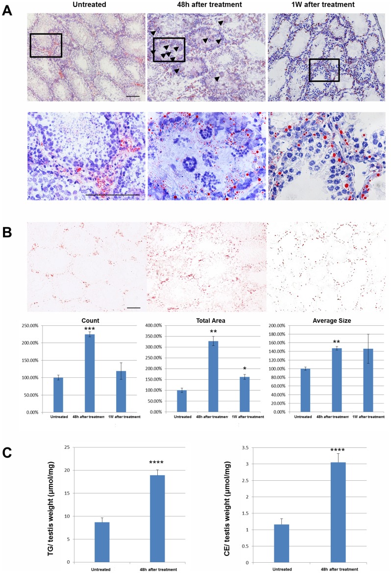
Figure 2. Morphological examination of lipid droplets was performed by oil red O staining (red) and counterstaining using hematoxylin (blue).
A high magnification image of regions in the black rectangle is shown in the lower panels. Black triangles point to degenerated and apoptotic germ cells (A). Oil red O staining was quantified by counting number, total area, and average size of staining from discontinuous slides, using three samples per group (B). Bars represent the mean ± SEM, Scale bar = 50 µm. (C) The lipid composition (cholesterol ester [CE] and triglyceride [TG]) in the droplets of testes from the untreated and the 48 hours after treatment groups. (*P<0.01, **P<0.001, ***P<0.0001, ****P<0.00001 compared to the untreated control).

