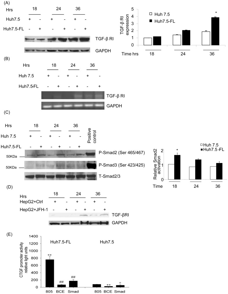
Figure 6. Expression of CTGF in Huh7.5-FL cells is Smad-dependent.
Cell lysates from Huh7.5 or Huh7.5-FL cells were collected at different time points and blotted with anti-TGF-βRI (A) or phospho-Smad 2, phospho-Smad3 and total Smad2/3 antibodies (C). The bar graphs show the quantitative analyses of TGF-β RI and p-smad2 protein expression as obtained by densitometry. RNA from Huh7.5 or Huh7.5-FL cells was used in the reverse transcriptase PCR to analyze the TGF-β RI expression (B). (D) HepG2 cells were transfected with and without JFH-1 RNA. The cell lysates were collected at different time points and blotted for TGF-βRI. (E) Huh7.5 and Huh7.5-FL cells were transfected with different CTGF promoter/SEAP reporter constructs for 48 hrs. CTGF promoter activity was determined by measuring SEAP reporter expression. ** P<0.001 versus Huh7.5 cells; + P<0.05 versus Huh7.5 cells; and ## P<0.001 versus Huh7.5-FL cells. Data represent mean ± SD of 3 independent experiments.

