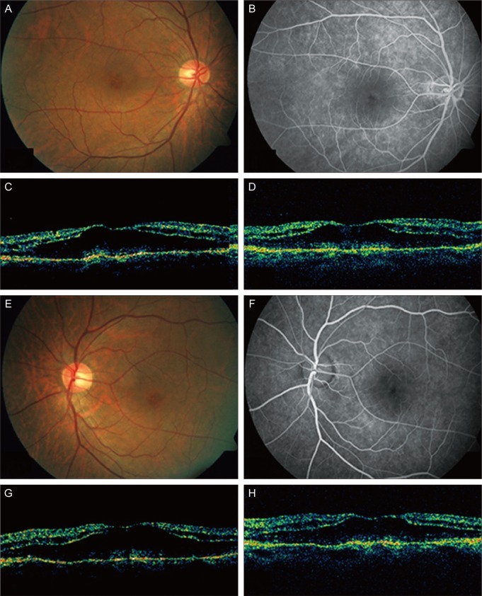
Fig. 1.
(A-D) The right eye of the patient. (A) Before intravitreal bevacizumab plus triamcinolone injection, fundus showed cystic change of the macula. (B) Fluorescein angiograms (FA) showed hyperfluorescence in the macula in the late frame. (C) Optical coherence tomography (OCT) showed cystoid macular edema (CME). (D) Six weeks after the injection, CME was unchanged in OCT. (E-H) The left eye of the patient. (E) Before intravitreal bevacizumab plus triamcinolone injection, fundus showed cystic change of the macula. (F) FA showed hyperfluorescence in the macula in the late frame. (G) OCT showed CME. (H) Six weeks after the injection, CME was unchanged in OCT.

